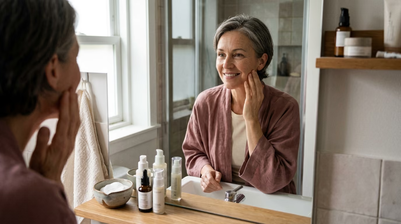

Skin aging involves complex biological changes across multiple tissue layers, driven by cellular decline, hormonal shifts, and long-term environmental exposure.





Most skincare marketing treats skin aging as a simple surface defect that can be smoothed away with a single topical cream. This surface-only narrative misrepresents the true biology of human skin. Skin aging is not a uniform or superficial event. It is a multi-layered, biological restructuring that involves bone density, subcutaneous fat, structural proteins, hormonal signaling, and cellular repair systems.
Understanding how skin changes over time requires looking beneath the visible surface. When you examine the biological mechanisms that drive tissue changes, you can make informed, rational choices for skin longevity and healthy aging. This guide examines the structural, biochemical, and environmental factors that govern how skin matures over the human lifespan.
Skin appearance is the direct physical expression of several interconnected anatomical layers. When visible changes occur at the surface, they reflect structural alterations occurring across the entire tissue stack.
The outermost layer of the skin is the stratum corneum, which serves as the primary barrier against environmental hazards and moisture loss. In youthful tissue, keratinocytes regenerate efficiently and progress through an orderly maturation cycle that lasts roughly 28 days. As chronological aging progresses, epidermal cell turnover slows considerably. This delay leads to an accumulation of irregularly shed surface cells, which impairs optical light reflection and makes the complexion look dull.
The lipid matrix of the stratum corneum also changes with age. Natural production of ceramides, cholesterol, and free fatty acids declines, while natural moisturizing factor levels diminish. These changes weaken the epidermal permeability barrier. As a result, mature skin experiences increased transepidermal water loss and heightened susceptibility to irritants.
True skin dryness is fundamentally a barrier lipid deficiency rather than simple dehydration. When the lipid matrix is depleted, the epidermis struggles to retain water even when systemic hydration is adequate. This barrier vulnerability also prolongs the time required for the surface to recover after physical trauma, chemical peeling, or environmental irritation.
Between the outer epidermis and the deeper dermis lies the dermal-epidermal junction. In youthful tissue, this interface is shaped into deep interdigitations known as rete ridges and dermal papillae. These microscopic folds provide mechanical anchoring and create a vast surface area for the diffusion of oxygen, micronutrients, and signaling molecules between vascularized dermis and avascular epidermis.
One of the most significant structural changes in mature skin is the flattening of this junction by more than 33 percent. As the interlocking ridges recede, the surface area connecting the two layers diminishes. This architectural flattening weakens mechanical resistance against shearing forces, which makes older skin significantly more prone to tearing, blistering, and physical injury.
The loss of contact area also impairs molecular communication and nutrient delivery to the basal epidermis. Keratinocyte stem cells receive fewer growth signals and fewer metabolic building blocks. This microvascular and structural deficit directly contributes to delayed wound healing, persistent thinning of the overlying epidermis, and reduced tissue resilience under physical stress.
The dermis provides the bulk of the skin's mechanical strength, elasticity, and hydration. This layer consists primarily of an extracellular matrix synthesized by specialized cells called fibroblasts. Collagen fibers, particularly Type I and Type III collagen, make up roughly 80 percent of the dry weight of the dermis and provide tensile strength. Interwoven among these collagen bundles is a network of elastin and fibrillin fibers that allow the tissue to stretch and return to its original shape.
With advancing age, the balance between matrix synthesis and matrix degradation shifts unfavorably. Fibroblasts become less active, synthesizing fewer new structural proteins while producing higher baseline levels of matrix-degrading enzymes. The collagen scaffolding becomes progressively fragmented, disorganized, and sparse. This degradation reduces dermal thickness, leading to visible skin laxity, fine lines, and a characteristic crepey texture.
In chronically sun-exposed tissue, elastic fibers undergo a severe structural alteration known as solar elastosis. Instead of a delicate, functional network of recoil fibers, damaged elastotic material accumulates into disorganized, non-functional protein masses. This abnormal remodeling does not restore youthful elasticity. Instead, it creates stiff, inelastic skin that deepens into permanent mechanical wrinkles when subjected to repeated facial expressions.
Understanding the complexity of collagen and structural matrix aging shows why structural aging cannot be solved by simply stimulating superficial cell turnover.
Beneath the dermis lies the subcutaneous fat layer, organized into distinct superficial and deep adipose compartments. These fat pads provide volume, contour, and smooth transitions across the facial skeleton. Facial aging involves compartment-specific changes, where some fat pads atrophy while others shift downward under the influence of gravity and ligamentous laxity.
As deep fat compartments lose volume, the overlying dermal envelope loses its structural foundation. This volumetric loss creates hollows in the tear troughs, temples, and mid-face, while downward migration of superficial fat contributes to nasolabial folds and jowling along the jawline. At the same time, fibrous connective tissue surrounding adipocytes thickens, reducing tissue pliability.
Beneath the adipose tissue, structural facial bones undergo continuous remodeling and gradual resorption. Bone loss around the orbital rim, pyriform aperture, and mandible reduces the underlying framework that supports soft tissues. Many visible folds and sagging contours that are casually blamed on skin surface aging are actually caused by changes in these deeper adipose and skeletal layers.
The structural decline of skin layers is governed by specific cellular and biochemical pathways. Chronological aging, oxidative stress, immune signaling, and metabolic byproducts continuously alter cellular behavior.
Dermal fibroblasts require physical tension to maintain their structural synthesis. In young, organized skin, fibroblasts attach firmly to dense collagen bundles, which stretches the cell body and stimulates normal gene expression for Type I collagen and hyaluronic acid. When matrix metalloproteinase enzymes break down surrounding collagen, fibroblasts lose their mechanical anchors.
Without mechanical tension, fibroblasts collapse into a rounded, inactive morphology. These uncoupled cells drastically downregulate collagen synthesis and increase their production of catabolic enzymes, which further damages the surrounding matrix. This process creates a self-sustaining cycle of matrix destruction and cellular shutdown.
Over time, an increasing proportion of fibroblasts enter cellular senescence. Senescent cells permanently cease division but remain metabolically active, secreting a destructive mixture of pro-inflammatory cytokines, chemokines, and proteases. This secretome, known as the senescence-associated secretory phenotype, degrades adjacent extracellular matrix and drives nearby healthy fibroblasts into premature dysfunction.
Matrix metalloproteinases, or MMPs, are zinc-dependent endopeptidases responsible for remodeling the extracellular matrix. Under healthy conditions, enzymes like collagenase (MMP-1), gelatinase (MMP-2 and MMP-9), and stromelysin (MMP-3) clear away damaged structural proteins during tissue repair. When cellular stress becomes chronic, MMP production overwhelms natural tissue inhibitors.
Low-grade, persistent systemic inflammation, often termed inflammaging, keeps MMP concentrations elevated for years. Chronically elevated cytokines such as interleukin-1, interleukin-6, and tumor necrosis factor-alpha continuously stimulate MMP gene expression. This chronic enzymatic activity quietly dissolves healthy structural scaffolding long before visible wrinkles appear on the surface.
Environmental exposures amplify this inflammatory cascade. Solar ultraviolet radiation triggers cell-surface cytokine receptors within minutes of exposure, sending signals that flood the dermis with MMP-1. This process breaks down intact collagen fibers and leaves fragmented collagen fragments that permanently alter dermal density.
Hormones exert a strong regulatory influence on cutaneous physiology, with estrogen playing a central role in maintaining dermal thickness and hydration. Estrogen receptors located on dermal fibroblasts and epidermal keratinocytes directly stimulate the production of Type I and Type III procollagen, tropoelastin, and fibrillin. Estrogen also supports the synthesis of dermal glycosaminoglycans, which maintain tissue turgor and water retention.
During the perimenopausal and postmenopausal transitions, circulating estradiol levels decline precipitously. This hormonal withdrawal alters skin metabolism. Clinical evaluations demonstrate that women lose approximately 30 percent of their cutaneous collagen within the first five postmenopausal years, accompanied by an average collagen loss of roughly two percent per year thereafter.
The loss of estrogenic signaling also compromises the epidermal barrier. Sebum production decreases, epidermal cell renewal slows, and glycosaminoglycan synthesis falls, reducing dermal moisture retention. These changes explain why many women experience sudden dryness, increased skin fragility, accelerated tissue laxity, and pronounced fine wrinkling during midlife.
Glycation is a spontaneous, non-enzymatic chemical reaction between circulating reducing sugars and structural amino groups on proteins, lipids, and nucleic acids. This initial reaction forms unstable Schiff bases and Amadori products, which slowly undergo complex rearrangements to form irreversible advanced glycation end products, commonly abbreviated as AGEs.
Long-lived structural proteins with slow turnover rates, such as dermal collagen and elastin, are especially vulnerable to glycation. When AGEs accumulate on collagen fibrils, they form rigid, covalent cross-links between adjacent protein strands. This cross-linking prevents collagen fibers from sliding smoothly past one another, making the skin matrix brittle, stiff, and prone to micro-cracking under mechanical stress.
Beyond altering physical tissue mechanics, AGEs bind to specific cell-surface receptors known as RAGE. Activation of RAGE on fibroblasts, keratinocytes, and endothelial cells triggers intracellular signaling cascades that generate reactive oxygen species and activate inflammatory transcription factors. This receptor-driven signaling suppresses normal collagen synthesis, accelerates MMP expression, and impairs microvascular blood flow, which hinders tissue repair and contributes to a dull, sallow complexion.
Mitochondria generate the cellular energy required for protein synthesis, barrier lipid formation, and DNA repair. During oxidative phosphorylation, mitochondria naturally produce reactive oxygen species as metabolic byproducts. Under normal conditions, endogenous antioxidant enzymes such as superoxide dismutase, catalase, and glutathione peroxidase neutralize these oxidants before they damage cellular components.
With chronological aging, mitochondrial DNA accumulates mutations due to its close proximity to reactive oxygen generation and its lack of protective histone proteins. These mutations impair the electron transport chain, creating a dysfunctional state where mitochondria produce fewer ATP molecules and significantly more reactive oxygen species. This energy deficit leaves cells unable to perform baseline repair functions.
When intracellular oxidants overwhelm antioxidant defenses, oxidative stress damages cell membranes through lipid peroxidation, breaks nuclear DNA strands, and cross-links cellular proteins. UVA radiation amplifies this damage by penetrating deep into the dermis and generating singlet oxygen and hydroxyl radicals. This oxidative cascade damages fibroblasts, degrades the extracellular matrix, and accelerates cellular senescence across all cutaneous compartments.
Separating genuine dermatological science from commercial promotion requires a rigorous examination of published clinical studies, randomized controlled trials, and epidemiological data.
The most definitive evidence regarding environmental skin aging comes from a landmark randomized controlled trial conducted by Green and colleagues in Queensland, Australia. The trial evaluated 903 adults over a four-and-a-half-year period to assess the effect of daily broad-spectrum sunscreen application compared to discretionary sunscreen use.
The study utilized standardized microtopography scoring of skin surface impressions to objectively measure cutaneous micro-anatomy and aging progression. The data revealed that participants assigned to daily broad-spectrum sunscreen use experienced 24 percent less skin aging progression over the study period compared to the discretionary-use control group (relative odds ratio of 0.76; 95 percent confidence interval, 0.59 to 0.98).
The trial demonstrated that daily application prevented measurable photoaging progression across both young and middle-aged adults. However, the same trial found that oral beta-carotene supplementation had no statistically significant effect on skin aging scores, demonstrating that dietary antioxidant supplements cannot replace direct topical protection.
Epidemiological research confirms the dominant contribution of ultraviolet radiation to visible facial wrinkling. A systematic review evaluating environmental risk factors established that individuals with more than one hour of daily recreational or occupational sun exposure had nearly double the odds of developing severe facial wrinkling compared to unexposed cohorts (pooled odds ratio of 1.90; 95 percent confidence interval, 1.14 to 3.18), after adjusting for tobacco use and chronological age.
While public health messaging historically cited estimates suggesting that sun exposure accounts for up to 80 percent of visible facial aging, actual clinical outcomes vary across demographics. The biological impact depends heavily on baseline melanin pigmentation, cumulative UV dose, geographic latitude, and personal photoprotective habits.
Clinical and epidemiological investigations have linked airborne particulate pollution to accelerated skin aging, particularly pigmentary abnormalities. In extensive cohort studies, exposure to fine particulate matter (PM2.5 and PM10), traffic-related soot, and nitrogen dioxide showed a direct statistical correlation with increased solar lentigines and deeper nasolabial folds.
Mechanistic studies indicate that polycyclic aromatic hydrocarbons bound to airborne particles penetrate the stratum corneum and bind to the aryl hydrocarbon receptor in keratinocytes and melanocytes. This receptor activation increases intracellular reactive oxygen species and upregulates tyrosinase activity, which drives irregular melanin synthesis and causes persistent dyschromia and lentigines.
Clinical biopsy data and ultrasound imaging studies have documented the exact timeline of postmenopausal dermal thinning. Observational data indicate that cutaneous collagen density decreases by approximately 2.1 percent per postmenopausal year, while skin thickness decreases by roughly 1.1 percent annually.
Systemic hormone replacement therapy studies have shown partial preservation of dermal thickness and collagen content in postmenopausal women. However, hormone therapy carries systemic health risks and is not indicated solely as a cosmetic treatment. Topical estrogens and selective estrogen receptor modulators continue to be researched, but systemic endocrine treatments require careful medical oversight.
While skin longevity research has advanced considerably, significant scientific limitations remain. Acknowledging what the data does not prove protects against exaggerated clinical claims.
Dermatological research often uses surrogate biomarkers to quantify biological skin age. Tools such as skin autofluorescence devices measure cutaneous AGE accumulation, while cutometers measure mechanical skin elasticity and suction recovery. Transepidermal water loss meters evaluate barrier integrity, and high-frequency ultrasound measures dermal thickness.
While these tools provide valuable data, no single biomarker accurately captures the complete biological state of human skin. A person may exhibit high skin autofluorescence due to dietary habits while maintaining excellent dermal thickness and minimal photoaging. Conversely, severe photoaging can exist alongside normal barrier recovery metrics. Clinicians and consumers must avoid treating any single diagnostic measurement as an absolute index of biological age.
A common limitation in beauty science research is the over-reliance on isolated cell cultures and animal models. In vitro experiments often expose cultured fibroblasts to high concentrations of botanical extracts, peptides, or growth factors, reporting massive increases in Type I procollagen expression.
These laboratory results rarely translate directly to human skin. In a living human, an applied molecule must penetrate an intact stratum corneum, survive enzymatic degradation in the epidermis, and reach the target fibroblasts in the dermis at a biologically active concentration. Many heavily advertised cosmetic ingredients show impressive results in cell cultures but fail to produce measurable changes in controlled human clinical trials.
Much of the foundational literature on photoaging and structural decline was conducted on fair-skinned individuals of European ancestry (Fitzpatrick phototypes I through III). These populations exhibit a high vulnerability to ultraviolet-induced erythema, early collagen fragmentation, and deep wrinkling.
These historical findings cannot be applied uniformly to more pigmented skin phenotypes (Fitzpatrick phototypes IV through VI). In more darkly pigmented individuals, higher baseline melanin content provides intrinsic photoprotection against UV-induced collagen breakdown. Visible aging in pigmented skin often presents 10 to 20 years later and is dominated by post-inflammatory hyperpigmentation, uneven tone, and structural fat redistribution rather than fine surface wrinkling. Clinical recommendations must account for these distinct ethnic and structural differences.
To make sound skincare decisions, you must be able to distinguish scientifically verified physiological realities from persuasive marketing narratives.
Translating skin biology into a practical routine requires focusing on interventions supported by rigorous clinical evidence. A successful strategy addresses barrier preservation, daily photoprotection, targeted topical signaling, and metabolic health.
Because ultraviolet radiation is the primary modifiable driver of premature skin aging, daily broad-spectrum sun protection is the foundation of any evidence-based routine. Photoprotection should be maintained consistently throughout the year, regardless of cloud cover or season.
For an extensive review of how external stressors degrade tissue structure, consult our guide on lifestyle, recovery, and environmental aging.
Maintaining a healthy epidermal barrier prevents transepidermal water loss, calms inflammatory signaling, and protects deeper dermal matrix proteins from external irritants.
A comprehensive look at maintaining foundational skin health is detailed in our guide to skin health and physiology.
Only a small number of topical active ingredients possess robust clinical data proving they can stimulate collagen synthesis and repair extracellular matrix structure.
To understand the biochemical validation behind these and other active ingredients, explore our analysis of beauty science and clinical skincare.
Because advanced glycation end products stiffen structural proteins and drive continuous low-grade inflammation, metabolic health directly influences skin longevity.
For deeper insights into how dietary patterns influence skin structure and cellular longevity, review our resources on nutrition and skin health.
Systemic lifestyle factors influence the balance between cellular damage and nocturnal tissue repair.
Chronological aging is an intrinsic biological process characterized by cellular senescence, slower cellular renewal, and gradual thinning of the epidermis and dermis. The skin remains structurally smooth and unblemished, with fine wrinkles and mild laxity. Photoaging is an accelerated extrinsic process caused by ultraviolet radiation. It results in profound matrix disorganization, deep leathery wrinkles, severe solar elastosis, rough texture, and irregular pigmentation.
Over-the-counter topical products cannot fully reverse the systemic hormonal collagen loss that occurs during menopause. However, an evidence-based routine combining topical retinoids, L-ascorbic acid, barrier-repair lipids, and daily broad-spectrum sunscreen can stimulate local procollagen synthesis, slow matrix degradation, and noticeably improve skin thickness, elasticity, and hydration.
Matrix-degrading enzymes are active throughout life as part of normal tissue remodeling. However, around the mid-twenties, baseline collagen production begins declining by roughly one percent per year, while environmental insults like UV exposure and pollution accelerate enzyme activity. Establishing gentle barrier support and consistent photoprotection early in adulthood helps prevent premature structural breakdown.
Skin tears and bruises more easily with age due to two key structural changes: the flattening of the dermal-epidermal junction and the thinning of the dermis. The flattening of the junction reduces mechanical anchoring between skin layers, making the surface vulnerable to shearing tears. Simultaneously, dermal thinning and microvascular fragility reduce the protective cushioning around small blood vessels, allowing minor impacts to rupture capillaries and cause bruising.
Once advanced glycation end products form stable cross-links on dermal collagen and elastin, they are exceptionally difficult for the body to break down. Because cross-linked structural proteins resist normal enzymatic turnover, the most effective approach is prevention. Maintaining stable blood glucose levels, consuming a nutrient-dense diet, avoiding smoking, and using daily sun protection helps minimize the formation of new cross-links over time.
Taking a measured, science-based approach to skin health allows you to make informed decisions that support structural integrity and tissue resilience over a lifetime.
Stay connected for research and practical guidance on skin, hair, collagen, nutrition and beauty longevity. Clear ideas for people who want to understand how appearance changes with age and make better-informed choices over time.


Understand your skin, hair and body better without chasing every new trend, treatment or promise.
explore the Blog