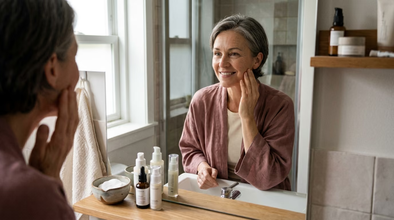

Dermal matrix architecture undergoes structural collagen fragmentation over time, altering fibroblast function and driving intrinsic and photoaging skin.





Most discussions about skin aging treat collagen like water in a leaking reservoir. The popular narrative suggests that our bodies simply run out of collagen over time, and that refilling the reservoir is all it takes to restore youthful skin.
Biological reality is far more complex and interesting. Skin aging is not just a passive decline in the amount of collagen your cells produce. It is a systemic breakdown in the mechanical communication between dermal cells and the surrounding structural matrix.
As the structural scaffolding of the skin degrades, the cells responsible for building new scaffolding lose their physical support. This loss of tension causes the cells to collapse, which triggers them to produce even fewer structural proteins and more tissue-degrading enzymes.
Understanding how the dermal matrix changes over time requires looking past simplified marketing slogans. By examining the true biology of extracellular matrix remodeling, we can separate genuine interventions from superficial claims and make informed decisions about skin longevity and healthy aging.
The dermis is the thick, supportive connective tissue layer located directly beneath the epidermis. It provides structural strength, elasticity, and nutritional support to the skin.
Collagen is the primary structural component within this layer. In fact, collagen accounts for roughly 90 percent of the dry weight of human skin. It is the main load-bearing element that allows skin to resist mechanical stress, deformation, and tearing.
The extracellular matrix, often abbreviated as the ECM, is the complex three-dimensional network surrounding all dermal cells. This environment is not an inert filler material. It consists of fibrillar proteins, elastic fibers, structural glycoproteins, proteoglycans, and glycosaminoglycans.
Scientists refer to the collection of structural genes and proteins in this network as the core matrisome. In human skin, the core matrisome includes 43 collagen subunits, 35 proteoglycans, and approximately 200 glycoproteins.
The major structural collagens in the dermis belong to distinct families, each performing specific mechanical duties:
Type I collagen is the most abundant protein in adult human skin. It forms thick, dense fibril bundles that deliver high tensile strength. Research shows that age-related reductions in type I collagen are the primary driver of dermal thinning and structural fragility.
Type III collagen forms narrower fibrils that provide compliance, suppleness, and structural flexibility. It is abundant in young tissues, vascular walls, and active repair sites. Together, type I and type III collagens form the bulk of the dermal structural scaffold.
Type V collagen intercalates with type I collagen to regulate the initiation and diameter of new collagen fibrils. While it represents a smaller quantitative fraction of the dermis, it is essential for proper fibril assembly.
Skin also contains specialized collagens that anchor different tissue layers together. Basement membrane collagens, such as type IV, and anchoring collagens, such as type VII, secure the dermal-epidermal junction. Without these specialized structural bridges, the upper skin layers can separate more easily under physical stress.
Surrounding these collagen fibers is an amorphous ground substance rich in proteoglycans and glycosaminoglycans. Proteoglycans such as decorin, biglycan, and versican attach to collagen fibrils to regulate their spacing and hydration. Sulfated glycosaminoglycans hold water molecules within the matrix, creating turgor pressure that helps the dermis absorb mechanical compression.
Dermal fibroblasts are the specialized cells responsible for synthesizing, organizing, and remodeling the extracellular matrix. For decades, popular beauty media portrayed fibroblasts as simple, passive factories that churn out collagen on demand. Modern cell biology reveals that fibroblasts are highly sensitive sensory cells that continuously evaluate their physical surroundings.
Fibroblasts create collagen through a multi-step biosynthetic pathway. Inside the cell, ribosomes synthesize polypeptide chains that wind together into a triple-helix precursor called procollagen.
The fibroblast secretes procollagen into the extracellular space. There, specific enzymes cleave the terminal globular ends to produce mature collagen molecules.
These individual collagen molecules self-assemble into microfibrils, which then organize into higher-order collagen fibers. Finally, an enzyme called lysyl oxidase catalyzes covalent bonds between adjacent molecules. This enzymatic cross-linking stabilizes the fibers and provides remarkable mechanical strength.
To maintain normal function, fibroblasts must physically anchor themselves to the matrix. They bind to intact collagen fibers through cell-surface receptors called integrins.
When attached to a taut, intact matrix, fibroblasts stretch out and generate internal cytoskeletal tension. This mechanical tension acts as a physical signal that tells the cell it is structurally supported. Under this physical tension, fibroblasts maintain normal gene expression for matrix production and cellular repair.
Matrix synthesis is heavily controlled by transforming growth factor beta, commonly known as TGF-beta. In healthy skin, TGF-beta binds to specific cell-surface receptors, primarily the TGF-beta type II receptor.
This binding activates intracellular signaling molecules called Smad2 and Smad3. These molecules join with Smad4 and enter the cell nucleus, where they switch on genes that code for type I collagen, type III collagen, decorin, and fibronectin.
At the same time, this pathway suppresses the production of tissue-degrading enzymes. This keeps dermal production and degradation in a healthy, dynamic balance.
The breakdown of dermal collagen is driven by a family of zinc-dependent enzymes known as matrix metalloproteinases, or MMPs. Fibroblasts, keratinocytes, and infiltrating immune cells all produce these enzymes. In young, healthy skin, MMP activity is tightly controlled by tissue inhibitors of metalloproteinases, known as TIMPs.
Matrix metalloproteinases break down the dermal scaffold through an orderly sequence of enzymatic steps:
As skin ages, this balanced clearing mechanism breaks down. Levels of several matrix-degrading enzymes, including MMP-1, MMP-2, MMP-3, and MMP-9, rise substantially in dermal tissue. However, levels of protective TIMPs do not increase proportionally. This creates sustained proteolytic pressure on the structural matrix.
Compounding this problem is the accumulation of non-enzymatic cross-links over time. Environmental stressors, lipid peroxidation, and reactions with reducing sugars create stable, abnormal cross-links between adjacent collagen molecules.
These abnormal cross-links make fragmented collagen resistant to complete enzymatic removal. Instead of being cleared efficiently, broken collagen pieces linger within the tissue.
Type I collagen in human skin has an exceptionally long natural lifespan. Researchers estimate the biological half-life of dermal collagen to be approximately 15 years. Because turnover is so slow, structural defects, chemical modifications, and mechanical micro-damage accumulate across decades.
The aging dermis does not just suffer from an absence of collagen. It becomes cluttered with a disorganized web of fragmented, stiffened, and dysfunctional structural remnants.
This progressive fragmentation directly impairs fibroblast function through a destructive feedback loop:
When collagen fragments, fibroblasts lose their physical anchor points. Without mechanical resistance from the matrix, the cells shrink, contract, and round up.
This cellular collapse turns off mechanical signaling pathways and downregulates TGF-beta receptors. The collapsed fibroblasts downregulate procollagen synthesis and dramatically increase their production of MMPs.
Matrix damage alters cell behavior, and altered cell behavior accelerates matrix damage. This self-reinforcing loop is a central driver of long-term collagen and structural aging.
To evaluate the dermal matrix accurately, we must separate chronological aging from environmental damage. Intrinsic aging, or chronological aging, represents the inevitable biological changes that occur over time across all areas of the body. Photoaging represents external damage caused primarily by repeated exposure to solar ultraviolet radiation.
Intrinsic aging progresses quietly in sun-protected skin, such as the inner upper arm or lower abdomen:
Photoaging accelerates these natural processes while adding distinct pathological changes:
Every exposed area of skin, including the face, neck, and hands, experiences both processes at the same time. Chronological aging sets the baseline rate of cellular decline, while lifetime sun exposure dictates the severity of matrix destruction.
Because of this overlap, clinical photographs of facial wrinkles cannot show chronological aging alone. Distinguishing intrinsic changes from photoaging prevents us from confusing preventable ultraviolet damage with inevitable biological aging.
Biomedical researchers have established precise molecular measurements for how the extracellular matrix changes in aged human tissue. These quantitative findings clarify why aging skin behaves differently under mechanical stress.
A landmark study examined human skin biopsies from individuals aged 80 and older and compared them directly with tissue from young adults aged 20 to 30. Using laser-capture microdissection combined with quantitative real-time RT-PCR, researchers measured gene expression within fibroblasts in their native tissue environment.
The analysis revealed that mRNA levels for the TGF-beta type II receptor were 59 percent lower in aged dermis compared to young dermis. Interestingly, expression of the TGF-beta type I receptor showed no significant change.
This 59 percent reduction in the type II receptor explains why aged fibroblasts struggle to produce matrix proteins, even when TGF-beta is present. The cells lose their surface antennas for growth factor signaling.
The same research analyzed interstitial ground substance components. Total sulfated glycosaminoglycans were roughly 40 percent lower in aged skin than in young skin.
Decorin was identified as the most abundant interstitial proteoglycan in the human dermis, followed by biglycan and versican. The combined loss of sulfated glycosaminoglycans and decorin reduces dermal hydration, impairs growth factor binding, and disrupts fibril spacing.
To understand whether fibroblast decline is purely genetic or driven by the matrix environment, researchers compared gene expression profiles. When fibroblasts from older donors were removed from damaged tissue and cultured in isolation, their expression of 42 collagen genes, 29 proteoglycan genes, and 157 glycoprotein genes matched those of young cells.
This shows that aged fibroblasts retain their genetic ability to produce matrix components. Their impaired performance in living skin stems largely from the collapsed, fragmented physical matrix around them.
The matricellular protein CCN1 offers further insight into how the matrix regulates cellular repair. Following carbon dioxide laser wounding, CCN1 expression surges 22-fold within 24 hours, peaking at a 31-fold increase at three days before returning to baseline by three weeks.
This rise coordinates the initial inflammatory phase and matrix-clearing stage before structural remodeling begins. In chronically aged tissue, dysregulated CCN1 signaling can lock fibroblasts into a persistent state of matrix degradation.
Topical creams and serums featuring collagen are staples of cosmetic marketing. Commercial advertisements often claim these products rebuild dermal architecture and restore lost structural proteins. However, physical chemistry and dermatological research show that intact collagen cannot penetrate the skin to reach the dermis.
The primary obstacle is the stratum corneum, the outermost protective barrier of the epidermis. Under the established 500 Dalton rule of dermatological pharmacology, a molecule must generally have a molecular weight under 500 Daltons to penetrate an intact epidermal barrier.
A complete, triple-helical type I collagen molecule has a molecular weight of approximately 300,000 Daltons. It is roughly 600 times too large to pass through the stratum corneum.
Even hydrolyzed collagen fragments used in topical serums typically range between 2,000 and 5,000 Daltons. Because of this size barrier, topical collagen sits on the surface of the skin.
Topical collagen products can still offer real, noticeable benefits to the surface of the skin:
Several clinical trials have evaluated multi-ingredient topical formulations containing collagen or collagen-related peptides:
A clinical study evaluated a serum containing pro-collagen lipopeptides, apple extract, creatine, and urea. Investigators noted an 11 percent increase in measured dermal density after one week, and 71 percent of participants showed reductions in visible fine lines.
Because this was a multi-ingredient product tested over a short period, the improvements likely reflected enhanced surface hydration and optical effects rather than rapid deposition of new dermal collagen fibers.
In another trial, a 3 percent topical triple-peptide complex improved wrinkle depth parameters by 10 to 19 percent compared to untreated baseline, and 13 to 28 percent compared to placebo.
Signal peptides are small enough to pass into upper epidermal layers, where they may stimulate cell signaling pathways. However, this biological response is fundamentally different from absorbing intact collagen into the dermis.
When a topical product improves the skin within a few days, that change is driven by surface hydration, barrier support, and optical plumping. While valuable for skin comfort and appearance, it should not be confused with true structural remodeling of the dermis.
Oral collagen supplements approach matrix biology from a different angle. Instead of attempting to push large, intact proteins through the skin barrier, oral supplements deliver hydrolyzed collagen peptides directly into the digestive tract.
During manufacturing, enzymatic hydrolysis breaks large collagen structures into small dipeptides and tripeptides, primarily prolyl-hydroxyproline and hydroxyprolyl-glycine. These short peptide sequences are absorbed intact across the intestinal wall into the bloodstream.
Ingested collagen peptides do not travel directly to the skin to plug into existing collagen fibers. Instead, researchers propose that these circulating peptides act as biological messenger molecules.
When fragments like prolyl-hydroxyproline reach dermal tissue, they bind to surface receptors on fibroblasts. This interaction signals to the cell that matrix breakdown has occurred, which encourages the fibroblast to increase its production of hyaluronic acid and structural proteins.
Additionally, emerging research suggests these peptides may influence M2-like macrophage activity and moderate low-grade inflammatory responses in the tissue.
A substantial body of clinical trials has examined oral collagen hydrolysates across different populations:
While these clinical findings are encouraging, the research has clear methodological limitations that call for careful interpretation:
Consuming hydrolyzed collagen peptides is safe and can provide modest, measurable improvements in skin elasticity and hydration for many individuals. However, oral supplements are supportive nutritional tools, not standalone solutions for structural tissue loss.
The market for healthy aging products is full of simplified claims that misinterpret extracellular matrix biology. Comparing common marketing claims against scientific evidence helps set realistic expectations.
Reality: Wrinkles are caused by changes throughout all facial layers. Facial aging involves thinning of the epidermis, loss of elastic fiber function, fat pad atrophy, bone resorption, repetitive muscle contraction, and gravity. Degraded collagen is an important structural factor, but it is only one component of a larger anatomical process.
Reality: The term collagen covers a diverse family of proteins with distinct structural roles and biological behaviors. In consumer products, the source, extraction method, peptide molecular weight profile, and processing determine how a formulation performs. An unhydrolyzed native collagen behaves very differently in the body compared to specific, low-molecular-weight bioactive dipeptides.
Reality: Clinical research has not found a direct, linear dose-response relationship for collagen supplements. Positive outcomes have been recorded at doses ranging from 1 gram to 10 grams daily. Flooding the digestive tract with excess peptides does not force fibroblasts to produce matrix proteins beyond their physiological capacity.
Reality: Cellular senescence involves a permanent loss of division, metabolic reprogramming, and the persistent secretion of inflammatory cytokines. Research shows that many aged fibroblasts are simply quiescent or physically collapsed due to a lack of mechanical support. When placed into a supportive, tensioned environment, many older fibroblasts regain their normal capacity to synthesize structural matrix proteins.
Reality: Skin hydration, dermal density, elasticity, and histologic collagen synthesis are distinct clinical endpoints. A product can increase stratum corneum water retention within hours through humectant mechanisms without altering procollagen gene expression. Measuring a change in moisture levels is not proof of structural remodeling in the dermis.
Supporting the dermal extracellular matrix over time requires a balanced, science-based approach that protects existing structural proteins while encouraging fibroblasts to maintain healthy synthesis.
Preventing the rapid, ultraviolet-induced surge of MMP enzymes remains the most effective way to preserve your dermal scaffold. Daily use of broad-spectrum sunscreen protects against UVA wavelengths that penetrate into the deep dermis. Preventing chronic, low-grade solar elastosis spares type I and type III collagen fibers from unnecessary enzymatic cleavage.
Topical retinoids, such as all-trans retinoic acid or retinol, are backed by decades of research for supporting matrix health. Retinoids bind to nuclear retinoic acid receptors in dermal cells, stimulating procollagen gene transcription while suppressing transcription factors like AP-1 that drive MMP production. Consistent use helps restore balance to matrix synthesis and degradation.
A compromised epidermal barrier triggers inflammatory cascades that release matrix-degrading enzymes in the underlying dermis. Supporting the stratum corneum with physiological lipids, including ceramides, cholesterol, and fatty acids, minimizes water loss and calms underlying stress signaling.
Fibroblasts require a reliable supply of specific amino acids, including glycine, proline, and hydroxyproline, along with essential micronutrients to assemble collagen triple helices. Vitamin C is a required cofactor for the prolyl and lysyl hydroxylase enzymes that stabilize collagen molecules. A balanced, protein-adequate diet provides the raw materials your body needs for tissue repair.
For readers seeking further practical insights, our detailed reviews in nutrition and beauty from within examine how dietary patterns shape tissue health over time.
Dermal fibroblasts assemble amino acids into procollagen triple-helix chains inside the cell before secreting them into the extracellular matrix. Outside the cell, specific enzymes trim the ends of these chains, allowing them to self-assemble into microfibrils. An enzyme called lysyl oxidase then creates stable chemical cross-links between the fibers, building a durable structural scaffold that gives skin its strength.
Natural collagen production begins to decrease gradually in early adulthood, typically starting in our mid-to-late twenties. The dermis loses roughly 1 percent of its collagen content per year through normal chronological aging. However, cumulative sun exposure, lifestyle habits, and metabolic health heavily influence the actual rate of loss in visible areas like the face and neck.
No. Complete collagen molecules and large hydrolyzed fragments are far too large to pass through the stratum corneum into the dermis. Topical collagen creams act as effective surface moisturizers and film-forming agents that improve hydration and smooth the look of fine lines, but they do not rebuild the underlying structural matrix.
Type I collagen is the main structural protein in adult skin, forming thick, dense fibril bundles that provide tensile strength and mechanical resistance. Type III collagen forms narrower, more flexible networks that give tissues suppleness and compliance. While both are essential for healthy skin, type I collagen makes up the vast majority of total dermal dry weight.
Matrix metalloproteinases, or MMPs, are enzymes that break down matrix proteins during normal tissue maintenance and repair. As skin ages, and especially after ultraviolet sun exposure, MMP levels rise while their natural inhibitors remain unchanged. This creates an imbalance that accelerates the fragmentation of structural fibers and disrupts normal fibroblast function.
Yes. Laboratory research shows that when aged, collapsed fibroblasts are removed from damaged tissue and placed in a supportive physical environment with adequate mechanical resistance, they can resume normal matrix production. Interventions that promote healthy tissue structure and restore cellular signaling help support active matrix maintenance.
No. Swallowed collagen is broken down during digestion into basic amino acids and small dipeptides or tripeptides. These small peptides are absorbed into the bloodstream, where they can act as chemical signaling molecules that encourage fibroblasts to produce their own structural proteins and hyaluronic acid. They do not travel directly to the skin as pre-formed fibers.
Maintaining healthy dermal architecture over time requires protecting the existing extracellular matrix from preventable environmental damage while supporting the natural signaling pathways that keep fibroblasts active.
Stay connected for research and practical guidance on skin, hair, collagen, nutrition and beauty longevity. Clear ideas for people who want to understand how appearance changes with age and make better-informed choices over time.


Understand your skin, hair and body better without chasing every new trend, treatment or promise.
explore the Blog