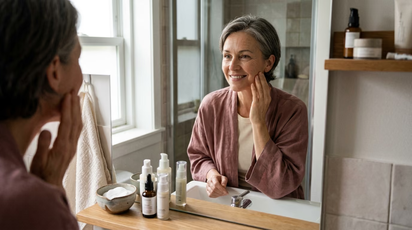

Three distinct tissue layers experience unique biological changes over decades, requiring targeted strategies to treat lines, sagging, barrier loss, and pigment concerns effectively.





You stand in front of the bathroom mirror under bright overhead lighting. You notice a fine web of lines around your eyes, a persistent dry patch near your cheekbone, and a shadow along your jawline that seems new. In response, you might purchase a brightening serum, a firming cream, and a strong exfoliating acid. A month later, your skin feels irritated, the dry patch is flaky, and the jawline shadow looks exactly the same.
This common frustration happens when we treat every visible change as a single, generic problem. Skin aging is not a uniform event. It is a complex collection of distinct biological processes occurring across different layers of tissue. A topical cream cannot lift descending fat pads. An exfoliating acid cannot calm dilated blood vessels.
Moving from reactive product shopping to informed skin planning requires a structured approach. By understanding the underlying biology of lines, folds, laxity, dryness, texture, redness, and pigment, you can match each visible change to the right level of care. This guide provides a taxonomy of skin aging concerns to help you set realistic priorities based on current scientific evidence.
The scientific understanding of cutaneous aging distinguishes between internal timelines and external damage. The core findings from clinical dermatology include:
To understand why different skin concerns require different solutions, we must look beneath the surface. Your skin consists of three primary layers: the epidermis, the dermis, and the subcutaneous tissue. Each layer undergoes distinct changes as decades pass.
The outermost layer, the epidermis, acts as a protective shield against environmental stressors and moisture loss. In younger skin, keratinocytes migrate from the basal layer to the stratum corneum in roughly 28 days. As intrinsic aging progresses, this turnover cycle slows down to 40 or 50 days. The slowed replacement of surface cells leads to a buildup of dull corneocytes, uneven light reflection, and rough texture.
Simultaneously, the epidermal synthesis of essential barrier lipids decreases. The lamellar bilayers of the stratum corneum require a precise ratio of ceramides, cholesterol, and free fatty acids to prevent water evaporation. When lipid production drops, transepidermal water loss rises. This dehydration causes the skin to lose pliability, causing fine crinkling across the surface.
Beneath the epidermis lies the dermis, a dense structural framework made of type I and type III collagen, elastin fibers, and glycosaminoglycans. Fibroblasts synthesize these structural components, while enzymes called matrix metalloproteinases break down old or damaged proteins.
In intrinsically aged skin, fibroblast activity naturally declines, producing less new collagen each year. Photoaging accelerates this decline drastically. Ultraviolet A rays penetrate deep into the dermis, generating reactive oxygen species. These free radicals activate the AP-1 transcription pathway, which triggers excessive production of matrix metalloproteinases.
Over time, collagen becomes fragmented, disorganized, and sparse. Elastin fibers degrade and clump into non-functional tangles, a condition known as solar elastosis. Without a robust dermal scaffold, the skin loses its resilience. The repeated folding of daily facial expressions gradually etches permanent static creases into the surface.
Aging does not stop at the dermal border. The subcutaneous layer contains discrete fat compartments, retaining ligaments, and underlying facial bones. Over time, facial fat compartments undergo selective atrophy and downward redistribution. The deep fat pads that support the midface lose volume, while superficial fat shifts lower.
At the same time, facial retaining ligaments loosen and bone resorption occurs around the eye sockets, cheeks, and jaw. These architectural changes cause the overlying skin to sag, creating deep shadows, hollows, and heavy folds. Because these issues originate in deep structural layers, topical products applied to the epidermis cannot resolve them. To read more about how these support networks function, review our guide on collagen and structural aging.
The dermatological literature offers strong evidence regarding what topical ingredients, energy devices, and lifestyle adjustments can achieve. Grounding your expectations in clinical data prevents wasted investment in unproven solutions.
The clearest evidence for slowing visible aging comes from a randomized controlled trial conducted in Nambour, Australia. The trial followed 903 adults under the age of 55 for 4.5 years. Participants were divided into two primary groups: one assigned to use broad-spectrum SPF 15 or higher sunscreen daily, and another using sunscreen at their own discretion.
The researchers assessed skin aging by analyzing microtopographic silicone impressions of the back of the hands at baseline and after 4.5 years. The results showed that the daily sunscreen group experienced 24% less skin aging than the discretionary group. Participants in the daily group showed no detectable increase in microtopographic skin aging over the entire study period.
This landmark trial confirms that photoprotection is a potent preventive intervention. However, the data also shows that sunscreen preserves existing skin architecture rather than restoring extensive past damage. Photoprotection serves as a shield against progressive breakdown rather than a complete reversal tool.
Topical retinoids represent the gold standard for evidence-based topical remodeling. Molecules such as tretinoin bind to retinoic acid receptors in skin cells, normalizing keratinocyte differentiation and stimulating dermal fibroblasts to produce new procollagen.
Systematic reviews of tretinoin clinical trials demonstrate visible improvements in fine wrinkles, mottled hyperpigmentation, and surface roughness. Clinical benefits often appear as early as 12 weeks, with progressive improvements continuing through 24 months of consistent application.
Over-the-counter retinol and retinaldehyde also show positive effects on collagen production and epidermal thickness, though they require conversion steps in the skin that make them milder and slower-acting than prescription tretinoin. You can learn more about ingredient efficacy across our beauty science articles.
For mild to moderate tissue laxity, energy-based technologies use heat to stimulate collagen contraction and neocollagenesis. Monopolar radiofrequency delivers thermal energy into the deep dermis while protecting the epidermis with contact cooling. The heat causes immediate denaturation of collagen fibrils, followed by a secondary healing response that creates new collagen over two to six months.
Microfocused ultrasound targets deeper tissue planes, including the superficial muscular aponeurotic system. Clinical evaluations show modest, measurable tightening of the jawline and submental area in selected candidates. However, the outcomes are gradual and subtle. Energy devices provide incremental structural support, not the dramatic transformation achieved by surgical repositioning.
While dermatology provides rigorous insights, consumers must interpret study claims with a critical eye. Many cosmetic marketing claims rest on flawed methodologies that do not reflect real-world usage.
A significant portion of published cosmetic literature relies on small pilot studies with 15 to 30 participants. These small samples make it difficult to detect subtle differences between active formulations and simple hydrating vehicles.
Furthermore, many studies run for only four to eight weeks. Because human collagen remodeling takes several months, short-term trials primarily measure the temporary plumping effects of hydration rather than permanent structural changes. When evaluating evidence, prioritize long-term, double-blind, randomized controlled trials.
Researchers often use surrogate markers, such as biopsies showing increased type I procollagen mRNA or ultrasound imaging measuring dermal density. While these biological markers confirm cellular activity, they do not always translate into dramatic visible improvements. A 15% increase in procollagen synthesis does not mean that 15% of your wrinkles will disappear.
Many commercial studies compare an active ingredient formulation against bare skin rather than an identical base formula without the active ingredient. This practice inflates the apparent success of the product. Simply applying a basic moisturizer improves barrier function, smooths rough texture, and minimizes fine dehydration lines.
During our detailed investigation into environmental aging, we tested how various lifestyle factors impact skin barrier recovery. It was fascinating to see the data clearly show that simple habits like sleep and basic hydration often outperform the most expensive topical treatments. This reinforced our commitment to emphasizing foundational health over product hype.
To make sensible decisions about your skin, you must classify your concerns accurately. The following taxonomy breaks down the seven primary categories of visible aging, identifying the clinical patterns, biological drivers, and most effective treatment tiers for each.
Fine lines are shallow indentations confined to the epidermis and upper papillary dermis. They typically appear in areas with thin skin, such as around the eyes, across the forehead, and around the mouth.
Fine lines generally fall into three distinct types:
The primary drivers of fine lines include UV-induced matrix degradation and stratum corneum dehydration. When the outer barrier lacks water, the epidermis loses elasticity and creases easily under mechanical stress.
Your first priority is restoring hydration with barrier-supportive moisturizers and preventing further UV damage with daily sunscreen. For persistent static lines, introducing a well-tolerated retinoid encourages new collagen synthesis over time. If dynamic expression is the dominant cause, neuromodulator injections offer temporary relaxation of the underlying muscle.
Deep wrinkles are pronounced furrows that sit deep within the reticular dermis. Folds, such as nasolabial lines and marionette shadows, are not surface defects. They are complex landmarks created by shifting facial fat, ligament laxity, and tissue descent.
Long-term ultraviolet exposure causes severe solar elastosis, in which functional elastic fibers are replaced by disorganized protein clumps. Combined with the loss of deep fat support, the skin sags forward and folds over natural anatomical anchor points.
Topical products cannot fill deep structural hollows or erase established folds. While prescription tretinoin can improve overall dermal quality, deep creases require structural interventions. Hyaluronic acid fillers or biostimulatory injectables like poly-L-lactic acid can restore lost volume. For substantial tissue descent, surgical procedures provide the only reliable, anatomically direct correction.
Laxity describes the loss of cutaneous firmness and structural recoil. It presents as jowling along the mandible, drooping upper eyelids, softening of the neck contours, and general tissue descent.
Skin laxity results from a multi-layer breakdown. In the dermis, the progressive degradation of elastin fibers impairs the skin's ability to snap back after stretching. Below the dermis, the retaining ligaments that tether skin to underlying facial bone gradually stretch and weaken.
Cosmetic creams marketed for "firming" or "lifting" provide mild surface tightening via film-forming polymers, but they do not alter structural tension. Noninvasive energy treatments, such as monopolar radiofrequency and focused ultrasound, can induce modest dermal tightening. When laxity is severe with significant excess skin, surgical intervention remains the gold standard.
Dryness is characterized by skin tightness, flaking, rough scaling, and an uncomfortable feeling of irritation. Crepiness refers to a thin, delicate, paper-like surface texture that crinkles easily when gently compressed.
The underlying mechanism is a breakdown in stratum corneum barrier function. As sebum production and ceramide synthesis decline with age, the lipid matrix surrounding corneocytes develops gaps. Moisture escapes rapidly into the surrounding air, leading to cellular dehydration.
Addressing dryness requires a systematic barrier repair approach. Gentle, non-foaming cleansers protect remaining lipids. Formulations containing physiological ratios of ceramides, cholesterol, and fatty acids help repair the protective barrier. Humectants draw water into the upper layers, while occlusives seal it in. For more guidance on supporting your skin through lifestyle measures, explore our lifestyle and recovery resources.
Rough texture is an uneven, bumpy, or coarse surface feel. It is often accompanied by dullness, where the skin lacks natural radiance and appears flat or gray under ambient light.
When desquamation slows, dead keratinocytes do not shed evenly. Instead, they accumulate in irregular patches on the stratum corneum. This uneven surface scatters incoming light rather than reflecting it evenly, robbing the skin of its natural luminosity.
Topical keratolytics, such as low-concentration lactic, mandelic, or glycolic acids, loosen the desmosomal bonds holding dead cells together. This process encourages smooth, uniform shedding. Retinoids normalize keratinocyte maturation from the basal layer upward. However, excessive exfoliation can damage the lipid barrier, leading to increased irritation and redness.
Cutaneous redness can take several forms, including transient flushing, persistent diffuse erythema, and discrete telangiectasias (spider veins) around the nose and central cheeks.
Vascular redness stems from vascular reactivity, chronic inflammation, and underlying conditions like rosacea. Long-term UV exposure degrades the perivascular collagen matrix that supports delicate capillaries. Without physical support, these vessels remain dilated, becoming visible through the thinned epidermis.
Redness requires gentle handling. Harsh exfoliating acids and strong vitamin C serums can worsen vascular reactivity. Azelaic acid provides anti-inflammatory benefits for diffuse erythema. For fixed telangiectasias, vascular lasers such as pulsed dye lasers target oxyhemoglobin directly, sealing the vessels without damaging surrounding tissue.
Hyperpigmentation presents as discrete solar lentigines (sun spots), diffuse mottled discoloration, melasma patches, or post-inflammatory marks following acne or injury.
Melanocytes produce melanin in response to UV radiation, visible light, and inflammatory mediators. In photoaged skin, chronically damaged melanocytes produce pigment erratically, resulting in concentrated clusters of melanin. Melasma involves a combination of UV exposure, visible light, hormonal fluctuations, and underlying vascular inflammation.
Managing pigmentation begins with rigorous, broad-spectrum sun protection. Tinted sunscreens containing iron oxides are essential for protecting against high-energy visible light, which triggers stubborn pigment in deeper skin tones. Tyrosinase inhibitors such as hydroquinone, azelaic acid, tranexamic acid, and kojic acid help regulate melanin production. For comprehensive guidance on healthy skin maintenance, visit our skin resource hub.
Creating an effective routine requires structuring your interventions in a logical hierarchy. Rather than trying every active ingredient at once, build your plan step by step to protect your skin barrier and track your progress clearly.
Tier 1 forms the essential base of every routine. Without a stable barrier and daily photoprotection, advanced treatments can cause irritation and worsen pigmentation.
Once your barrier is resilient and sun protection is a daily habit, introduce targeted active ingredients based on your primary skin concern.
When topical skincare reaches its biological limit, in-office procedures can address deeper dermal concerns and localized issues.
Surgical intervention represents the definitive approach for structural descent, significant tissue laxity, and excess skin.
Marketing trends frequently oversimplify dermatology. Let us clarify several widespread misconceptions with scientific evidence.
Reality: Topical skincare products work primarily within the epidermis and upper papillary dermis. Tissue sagging along the jawline results from stretched retaining ligaments, fat pad redistribution, and bone resorption deep beneath the skin.
While a rich cream can hydrate the surface and improve fine crinkliness, it cannot alter underlying structural anatomy. Addressing true laxity requires in-office energy devices or surgical lifting.
Reality: Stinging, burning, and tingling are signs of sensory nerve irritation and acute barrier compromise, not evidence of deep cellular repair.
When you damage the stratum corneum with aggressive actives, you trigger inflammatory pathways. This chronic low-grade inflammation accelerates collagen breakdown and worsens post-inflammatory hyperpigmentation, particularly in deeper skin tones. The most sustainable improvements come from consistent, non-irritating applications.
Reality: While many pigmented spots are benign solar lentigines or freckles, some dark lesions represent serious medical conditions, including actinic keratoses or melanoma.
Applying brightening creams or seeking cosmetic chemical peels for an undiagnosed changing spot delays vital medical care. Any lesion that is asymmetric, exhibits irregular borders, contains multiple colors, or changes rapidly in size should be evaluated by a board-certified dermatologist before beginning cosmetic treatments.
Dehydration lines typically appear as an ultra-fine, shallow, paper-like network across your cheeks and forehead. They often look worse in air-conditioned environments, during dry winter months, or immediately after washing your face with a foaming cleanser.
If applying a hydrating humectant and a rich ceramide moisturizer smooths the surface within an hour, dehydration is the main culprit. Structural wrinkles, by contrast, remain visible as defined creases even when the skin is fully hydrated.
Clinical trials show that daily sunscreen use stops the progression of photoaging and allows the skin to direct its natural repair resources toward maintaining tissue health.
While some study participants show slight improvements in surface microtopography over several years, sunscreen functions primarily as a preventive shield. It prevents further matrix metalloproteinase activation, but it will not completely erase deep, established creases on its own.
Intrinsic aging naturally lowers the activity of your sebaceous glands and decreases the synthesis of essential epidermal lipids, particularly ceramides. Additionally, the skin's natural hyaluronic acid content in the dermis and epidermis declines over time.
A light lotion that provided sufficient hydration in your twenties may not supply enough protective lipids in your forties or fifties. Upgrading to a richer cream containing physiological lipids helps restore the weakened barrier.
Patients with melanin-rich skin (Fitzpatrick skin types IV through VI) must approach light and laser treatments with caution. High-energy devices can stimulate melanocytes to produce excess melanin, leading to post-inflammatory hyperpigmentation that is often more noticeable than the initial concern.
If you pursue in-office treatments, seek a board-certified dermatologist experienced in treating pigmented skin. They can select safer wavelengths, such as long-pulsed Nd:YAG or low-fluence picosecond lasers, and properly prepare your skin with topical tyrosinase inhibitors.
A deep wrinkle is an etched crease rooted directly in the dermal matrix, caused by long-term collagen breakdown and repetitive muscle movement.
A fold is a deeper landmark created when skin, soft tissue, and fat sag forward and pool against underlying facial retaining ligaments. Treating a fold requires restoring lost volume or lifting descended tissue, whereas treating a wrinkle involves resurfacing the dermal layer.
Skin is dynamic and changes across different seasons, life stages, and hormonal transitions. Revisit this taxonomy whenever you notice new visible patterns, when your current routine stops delivering results, or before investing in clinical procedures.
By categorizing your skin concerns by their biological origins, you can build a targeted, realistic routine that supports healthy skin over the long term.
Stay connected for research and practical guidance on skin, hair, collagen, nutrition and beauty longevity. Clear ideas for people who want to understand how appearance changes with age and make better-informed choices over time.


Understand your skin, hair and body better without chasing every new trend, treatment or promise.
explore the Blog