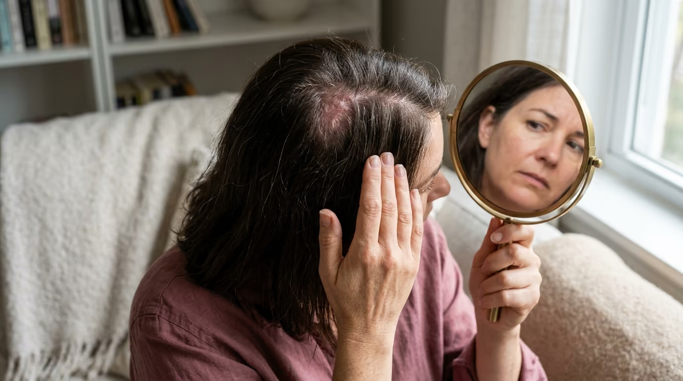

Routine shedding resolves naturally, but scarring alopecia causes permanent follicular damage, making early identification of redness, pain.





You part your hair in the mirror and notice a small area along your crown or hairline that looks slightly different than usual. The skin appears a bit smoother, perhaps mildly pink, or it feels slightly tender when you brush your hair. It is easy to dismiss these subtle scalp sensations as product buildup, dry skin, or routine age-related shedding. However, when hair loss involves inflammation that attacks the underlying follicular structure, waiting to see what happens can lead to permanent changes.
Recognizing the distinction between routine hair thinning and scarring alopecia is one of the most critical steps in scalp health. While many forms of hair loss temporarily pause the growth cycle, scarring disorders permanently destroy follicular architecture. Understanding the subtle early signs allows you to seek qualified medical evaluation while follicles remain viable.
Scarring alopecia, known clinically as cicatricial alopecia, represents a group of inflammatory disorders that target and destroy hair follicles. Once a follicle undergoes complete fibrosis, it loses the biological capacity to generate new hair fibers.
Scientific investigation into cicatricial disorders highlights several foundational principles:
Understanding these biological realities helps shift the focus from cosmetic concealment to targeted medical evaluation. Readers seeking comprehensive information on follicular preservation can review our hair growth and hair longevity resources for deeper context on long-term scalp care.
To understand why cicatricial alopecia causes permanent loss, one must examine the anatomy of the hair follicle. Deep within the upper portion of the follicle lies a specialized region called the follicular bulge. This bulge houses the epithelial stem cells responsible for regenerating the lower hair follicle during each new growth cycle.
In healthy scalp biology, hair follicles transition continuously through three distinct phases. The anagen phase represents active growth, catagen marks structural regression, and telogen serves as a resting phase before shedding. As long as the stem cell reservoir in the bulge remains intact, a new hair fiber can emerge. Even after extensive shedding, the follicle maintains its regenerative machinery.
In primary scarring alopecia, immune cells target this follicular bulge region directly. Depending on the specific condition, the inflammatory infiltrate consists primarily of lymphocytes or neutrophils. These immune cells release inflammatory mediators that break down the integrity of the follicular wall.
As inflammation intensifies, the sebaceous gland attached to the upper follicle is frequently destroyed early in the process. The epithelial stem cells within the bulge sustain irreversible cytotoxic injury. When these stem cells are eliminated, the follicle loses its capacity to re-enter the anagen growth cycle.
Following this inflammatory destruction, the body attempts tissue repair through fibroplasia. Fibroblasts migrate into the damaged follicular tract and deposit dense collagen fibers. This process converts the living follicular channel into a fibrous scar tract.
On the surface of the scalp, this internal scarring manifests as the loss of follicular openings. In healthy scalp tissue, pores or ostia are clearly visible under magnification. In scarred tissue, the skin becomes smooth and uniform because the underlying follicular conduit no longer exists.
Secondary scarring alopecia follows a slightly different biological pathway. Instead of an immune attack directed specifically at the follicle, external trauma destroys the tissue. Severe burns, radiation therapy, deep infections, or mechanical injury cause widespread dermal damage. The hair follicles are destroyed as collateral damage alongside surrounding connective tissue.
Regardless of whether the trigger is primary inflammation or secondary trauma, the final biological outcome is identical. Once scar tissue replaces the follicular stem cell niche, that specific follicle cannot regrow hair.
Clinical studies underscore the challenges and timelines associated with diagnosing scarring alopecias. Because these conditions develop gradually, patients and healthcare providers often mistake early stages for common conditions.
A retrospective cohort study examining 126 patients with lichen planopilaris revealed a mean interval of 33.9 months from initial disease onset to formal diagnosis. In that clinical group, 43 patients achieved complete disease remission during follow-up, while 83 did not. The researchers emphasized that persistent inflammation causes irreversible destruction. They concluded that closing this diagnostic gap is essential for preserving remaining hair.
Research exploring central centrifugal cicatricial alopecia highlights similar diagnostic complexities. In a retrospective chart review of affected patients, 79.5 percent exhibited hair loss concentrated at the vertex or crown. Scalp pruritus, or itching, was the most frequently documented symptom, occurring in 23.3 percent of patients. The remaining majority experienced either subtle tenderness, burning, or completely painless progression. This data demonstrates that the absence of severe pain does not rule out active disease.
Dermoscopic and trichoscopic research has yielded valuable diagnostic metrics. A specialized dermatoscopic study evaluated the presence of the peripilar white-gray halo in patients with central centrifugal alopecia. Researchers observed this specific halo in 38 out of 40 patients, representing a 95 percent prevalence rate. Another study confirmed this finding in 94 percent of patients, identifying it as a sensitive and specific marker of lamellar fibrosis around the outer root sheath.
Diagnostic accuracy varies across different trichoscopic markers and clinical subtypes. Comparative studies evaluating trichoscopy have demonstrated sensitivity rates of 100 percent for frontal fibrosing alopecia and lichen planopilaris in clinical settings. The diagnostic sensitivity for central centrifugal alopecia was documented at 66.7 percent in specific comparative cohorts. These figures demonstrate why trichoscopy is an exceptional tool for detecting early disease, though clinical biopsy remains the gold standard.
Investigating the scientific literature on hair science and scalp biology demonstrates that early inflammatory markers serve as vital guideposts. When medical evaluation occurs before extensive fibrosis takes place, therapeutic intervention can successfully halt ongoing destruction.
While scientific understanding of cicatricial alopecia continues to expand, existing research contains several important limitations that patients should understand.
Many published studies rely on retrospective cohort designs or single-center chart reviews. These studies gather data from patients who sought care at specialized academic dermatology centers. As a result, study populations often skew toward individuals with more advanced, severe, or treatment-resistant disease. These findings may not fully represent mild presentations managed in community settings.
Histopathological overlap presents another diagnostic challenge. Under microscopic examination, conditions such as lichen planopilaris, frontal fibrosing alopecia, and central centrifugal alopecia share strikingly similar features. All three exhibit lymphocytic inflammation, loss of sebaceous glands, and concentric perifollicular fibrosis. Pathologists must rely on careful clinicopathologic correlation, combining tissue slides with clinical scalp photographs, to reach an accurate diagnosis.
Furthermore, scalp examination findings can look markedly different across diverse skin tones and phototypes. In lighter skin phototypes, active inflammation frequently appears as bright pink or red perifollicular erythema. In deeper skin tones, active inflammation often manifests as subtle hyperpigmentation, violaceous discoloration, or mottled pigmentary alterations. Much of the older dermatological literature centered on lighter skin types, which historically led to under-recognition in darker phototypes. Modern research is addressing these gaps, but diagnostic criteria continue to evolve.
Finally, high-level randomized controlled trials evaluating therapeutic interventions for scarring alopecia remain scarce. Most clinical management guidelines are derived from observational studies, expert consensus, and case series. What the scientific literature proves definitively is that early suppression of inflammation prevents further follicular loss. However, evidence cannot yet predict with certainty which specific medication regimen will achieve remission in every individual patient.
Because cicatricial alopecia causes permanent loss once fibrosis occurs, recognizing early warning signs is vital. These signs often appear months or years before widespread baldness develops.
Scarring hair loss commonly begins as one or more localized patches of thinning. Unlike normal shedding, which occurs evenly across the entire scalp, scarring loss is often asymmetric or focal. A patch that slowly enlarges over several months deserves careful dermatologic evaluation rather than waiting for natural recovery.
In healthy scalp skin, hairs emerge cleanly from the surface. In active scarring alopecia, inflammation concentrates around the base of individual hair fibers. This creates a tiny collar of redness or a small tube of adherent white scale surrounding the hair shaft. These features may be difficult to see with the naked eye but become immediately apparent under magnification.
Scalp dysesthesia, which includes sensations of burning, stinging, itching, tenderness, or crawling, frequently accompanies active follicular inflammation. While general itching can occur with dandruff or dry skin, discomfort concentrated specifically within an area of thinning hair is an important warning sign.
Unexplained hair breakage near the scalp surface can serve as an early sign of central centrifugal alopecia. Before complete follicular destruction occurs, inflammatory damage weakens the structural integrity of the hair shaft within the follicle. If hairs continue snapping near the root despite gentle handling, an underlying inflammatory process may be present.
A gradual backward movement of the frontal and temporal hairline, accompanied by thinning or loss of the outer eyebrows, is characteristic of frontal fibrosing alopecia. The exposed scalp skin along the receded hairline often appears unusually smooth, pale, or shiny, lacking normal follicular pores.
Thinning that originates at the crown or vertex of the scalp and gradually expands outward in a circular pattern warrants attention. This centrifugal expansion is the classic presentation of central centrifugal cicatricial alopecia. Early stages may look like ordinary genetic thinning, but the presence of scalp tenderness or scale points toward a scarring process.
The appearance of small pimples, yellow crusting, bleeding, or fluid discharge around hair follicles indicates an active inflammatory or infectious condition. These symptoms are common in neutrophilic scarring disorders, such as folliculitis decalvans, and require rapid medical intervention.
As fibrosis develops beneath the surface, the physical texture of the scalp changes. The skin may feel unusually tight, bound down, or soft and spongy, depending on the subtype. Visual inspection may reveal areas that look porcelain white, shiny, or mottled with irregular brown or purple pigmentation.
Dermatologists classify cicatricial alopecias based on the primary type of immune cells driving the inflammation. Understanding these distinctions helps clarify why different conditions require unique clinical approaches.
Lichen planopilaris is a primary lymphocytic scarring alopecia that can develop in both women and men. It typically presents with scattered, irregular patches of hair loss accompanied by noticeable perifollicular scale and redness along the expanding borders.
Patients with lichen planopilaris often report substantial scalp itching, burning, and tenderness. When examined under trichoscopy, the active margins show distinct tubular scaling wrapping around hair shafts. Left unchecked, the patches can coalesce into larger areas of permanent hair loss.
Frontal fibrosing alopecia is considered a clinical variant of lichen planopilaris that exhibits a distinct anatomical pattern. It predominantly affects peri- and post-menopausal women, though it can occur in younger adults.
FFA characteristically produces symmetrical recession of the frontotemporal hairline, creating a distinct band of pale, smooth skin. Eyebrow loss, often beginning at the outer lateral edges, precedes or accompanies hairline recession in a large percentage of individuals. Small, isolated hairs, often termed lonely hairs, may persist within the band of receded skin.
Central centrifugal cicatricial alopecia is the most common form of primary scarring hair loss in women of African and Afro-Caribbean descent. It originates at the crown or vertex of the scalp and progresses outward in a roughly symmetrical, centrifugal fashion.
The underlying pathology involves chronic lymphocytic inflammation and lamellar fibrosis encasing the follicular root sheath. Patients may experience scalp tenderness, itching, or dull burning, though some remain completely asymptomatic during early stages. Central scalp hair breakage often serves as the initial clinical presentation.
Discoid lupus erythematosus is an autoimmune condition that can cause severe, localized scarring alopecia. DLE lesions frequently begin as red, inflamed, disc-shaped plaques covered by thick, adherent scale that extends into follicular openings.
As these lesions resolve, they leave behind pronounced central atrophy, permanent follicular loss, and marked pigmentary changes. Lesions often display white hypopigmentation in the center surrounded by a darker hyperpigmented border. Trichoscopy typically reveals tortuous branching blood vessels and large follicular keratotic plugs.
Unlike primary disorders where the follicle is the primary immune target, secondary scarring alopecia results from non-specific tissue destruction. External causes include:
Understanding these variations highlights why accurate professional classification is essential for selecting appropriate medical therapies.
Navigating a diagnostic evaluation for scarring alopecia involves several structured steps. Dermatologists specializing in hair disorders use clinical history, physical examination, trichoscopy, and pathology to establish a definitive diagnosis.
The evaluation begins with a detailed medical history. The clinician will ask when you first noticed shedding, thinning, or scalp discomfort, and whether the progression has been rapid or gradual.
Your history will cover daily hair-care practices, including heat tools, chemical relaxers, hair dyes, braids, extensions, and tight styling. A history of traumatic styling does not automatically mean hair loss is purely mechanical. In many cases, inflammatory scarring disorders coexist with routine styling practices.
The physician will also review personal and family histories of autoimmune diseases, thyroid disorders, skin conditions, and recent prescription medications.
The physical examination evaluates the entire scalp under bright, direct light. The physician examines hair density, part width, hair caliber, and the presence or absence of follicular openings across multiple zones.
Next, the dermatologist performs trichoscopy using a polarized dermatoscope. This handheld magnifying device illuminates the scalp surface, revealing details invisible to the naked eye.
Trichoscopy allows the clinician to identify:
Trichoscopy is also essential for selecting the optimal site for a scalp biopsy.
A scalp biopsy provides definitive histopathological confirmation when clinical and trichoscopic findings require verification. The procedure is performed in the office under local anesthesia.
The clinician typically takes one or two 4-millimeter punch biopsies. The choice of biopsy location is critical. Sampling the smooth, bald center of a scarred area yields only mature scar tissue, providing little diagnostic insight.
Instead, the biopsy must target the active inflammatory margin of the patch, where partially affected follicles and active immune cells reside. For suspected conditions like central centrifugal alopecia, clinicians often take two punches. One sample is sectioned horizontally to evaluate follicular density and cross-sectional architecture, while the second is sectioned vertically to assess inflammation depth.
Pathologists examine the tissue to determine whether the immune infiltrate is lymphocytic or neutrophilic, evaluate sebaceous gland destruction, and confirm the presence of concentric fibrosis.
Managing cicatricial alopecia requires long-term monitoring to confirm that disease activity has stabilized. Successful treatment is defined as stopping further hair loss rather than immediate regrowth.
Objective monitoring relies on several methods:
Because hair thinning has many causes, distinguishing cicatricial alopecia from non-scarring hair loss is an important clinical task.
Androgenetic alopecia, commonly known as female or male pattern hair loss, is driven by genetics and hormonal sensitivity. In this non-scarring condition, hair follicles undergo progressive miniaturization, producing progressively thinner, shorter hair fibers over successive cycles.
However, the follicular openings remain entirely intact, and stem cells within the bulge are preserved. The scalp surface looks normal, without localized scale, erythema, or structural scarring.
Telogen effluvium is an acute or chronic form of non-scarring hair shedding. It is typically triggered by systemic physiological stressors, such as severe illness, surgery, nutritional deficiency, or hormonal shifts.
Telogen effluvium produces generalized shedding across the entire scalp rather than localized, expanding patches. Follicular openings remain open and healthy, scalp symptoms are generally absent, and hair density routinely recovers once the underlying trigger resolves.
Alopecia areata is an autoimmune, non-scarring hair loss disorder. The immune system mistakenly attacks the hair bulb, arresting hair production and causing hairs to fall out in smooth, round patches.
Although alopecia areata can develop rapidly, the underlying follicle and its stem cells remain alive and structurally intact. Under trichoscopic evaluation, follicular openings remain clearly visible, often displaying yellow dots or exclamation-mark hairs. With appropriate treatment, complete hair regrowth remains biologically possible.
Traction alopecia stems from prolonged, repetitive mechanical tension applied to hair follicles by tight hairstyles, extensions, or heavy braids. In its early stages, traction alopecia is entirely non-scarring and reversible if tension is removed promptly.
However, if mechanical pulling persists over years, chronic inflammation around the follicle can induce permanent scarring. Recognizing when mechanical damage transitions into irreversible follicular loss requires professional evaluation.
Readers interested in the broader biological mechanisms of hair and skin health can review our beauty science articles for balanced, research-driven perspectives.
Not every hair concern requires emergency care, but unexplained hair loss should never be ignored. Use this three-tiered framework to determine the appropriate timeframe for medical evaluation.
Schedule a standard appointment with a board-certified dermatologist within several weeks if you notice:
Contact a dermatology clinic to request an expedited appointment within one to two weeks if you observe:
Seek prompt medical care within 24 to 48 hours at a clinic or urgent care facility if you experience:
Taking a proactive approach when you suspect an inflammatory hair disorder can protect healthy follicles. Here are practical, actionable steps to guide your care.
Before your medical visit, compile clear photographic documentation. Take well-lit, in-focus photographs of your hairline, temples, crown, and any specific areas of concern.
Repeat these photographs once a month under identical lighting conditions. Maintain a concise symptom log recording any episodes of itching, burning, tenderness, or unusual flaking. This objective record helps your dermatologist assess disease activity and progression speed accurately.
When scheduling, verify that the dermatologist has specific clinical experience in diagnosing and managing cicatricial alopecias. Bring a complete list of your current medications, dietary supplements, and relevant medical history.
Avoid washing your hair for 24 to 48 hours immediately prior to your examination. Refraining from shampooing preserves surface scale and inflammatory markers, making it easier for the clinician to identify active biopsy sites during trichoscopy.
While awaiting medical assessment, minimize physical and chemical trauma to your hair and scalp.
For further exploration of balanced wellness and lifestyle routines, visit our lifestyle and recovery guides.
Widespread myths about hair loss often delay appropriate medical care. Clarifying these misconceptions helps ensure that individuals take meaningful, evidence-based steps toward diagnosis.
Many people assume that destructive inflammation always causes intense pain, burning, or itching. In reality, multiple clinical studies demonstrate that scarring hair loss can progress silently.
A significant proportion of patients with central centrifugal alopecia or frontal fibrosing alopecia report zero pain or itching during early stages. Relying on physical discomfort as the sole indicator of disease can allow irreversible follicular loss to occur unnoticed.
Patients often expect a doctor to biopsy the most visible bald patch. However, taking a tissue sample from a completely smooth, hairless center is rarely helpful.
That tissue consists entirely of end-stage fibrous scar tissue, which cannot reveal the underlying immune mechanism. A skilled clinician will always biopsy the active margin of the patch, where early inflammation and partially affected follicles remain present.
When individuals notice their frontal hairline moving backward, they frequently assume it is ordinary androgenetic hair loss. As a result, many spend months or years applying over-the-counter hair-growth serums while the hairline continues to recede.
Frontal fibrosing alopecia specifically mimics genetic recession but requires entirely different medical therapies. The presence of eyebrow thinning, loss of follicular openings, or redness around remaining hairs points toward scarring rather than simple genetics.
Cosmetic marketing often suggests that botanical oils, scalp massages, or high-dose hair supplements can restore any form of thinning. In scarring alopecia, the biological regenerative machinery of the follicle has been permanently replaced by fibrous collagen.
No botanical oil, supplement, or cosmetic treatment can regenerate a destroyed stem cell bulge. Medical management focuses on saving the healthy follicles that remain, making early clinical intervention far more valuable than cosmetic remedies. You can read our broader editorial insights on hair and scalp health to learn how to distinguish reliable clinical evidence from unproven marketing claims.
Hair cannot grow back from follicles that have been completely destroyed and replaced by scar tissue. However, follicles that are only partially inflamed or structurally intact can be saved with prompt medical treatment. This biological reality is why dermatologists focus on early diagnosis and halting active disease progression.
In primary scarring alopecia, the hair follicle itself is the primary target of an inflammatory autoimmune process. In secondary scarring alopecia, the hair follicle is destroyed as collateral damage from an external injury, such as a severe burn, radiation therapy, deep infection, or mechanical trauma.
Treatment depends on whether the underlying inflammation is lymphocytic or neutrophilic. Dermatologists commonly utilize topical or intralesional corticosteroids, topical calcineurin inhibitors, oral anti-inflammatory antibiotics such as doxycycline, hydroxychloroquine, or other targeted immunomodulating medications to calm inflammation and prevent further loss.
No. While traumatic hair-care practices, excessive heat, and tight hairstyles can exacerbate scalp inflammation and traction, research indicates that CCCA is a complex, multifactorial medical condition. Genetic predispositions, intrinsic follicular structural variations, and chronic inflammation all play important roles in its development.
Progression rates vary substantially among individuals. Some forms, such as acute lichen planopilaris or folliculitis decalvans, can progress rapidly over several months. Other conditions, including frontal fibrosing alopecia, may develop slowly over several years with subtle periods of active inflammation alternating with relative quiescence.
Revisit this guide if you notice new scalp discomfort, an expanding area of hair thinning, unexplained hair breakage near the roots, or changes in your hairline or eyebrows. Keeping these clinical signs in mind ensures you can seek prompt dermatologic care whenever your scalp health changes.
Early recognition and specialized clinical evaluation remain your most effective tools for preserving long-term hair health and preventing irreversible follicular loss.
Stay connected for research and practical guidance on skin, hair, collagen, nutrition and beauty longevity. Clear ideas for people who want to understand how appearance changes with age and make better-informed choices over time.


Understand your skin, hair and body better without chasing every new trend, treatment or promise.
explore the Blog