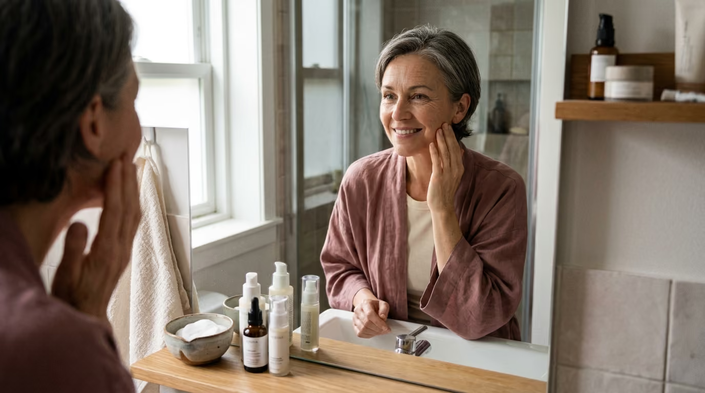

Daily exposure to sunlight and pollution triggers cellular oxidation and chronic inflammation, driving collagen loss, tissue damage, and premature skin aging.





Oxidative stress is not a simple accumulation of destructive free radicals, nor is inflammation merely an undesirable skin reaction. Both are fundamental biological processes that govern cellular signaling, tissue defense, and structural remodeling. When regulated appropriately, these pathways protect cells from pathogens, initiate wound healing, and stimulate adaptive resilience. When poorly controlled, prolonged, or overwhelmed by external exposures, they drive the structural and functional decline recognized as visible skin aging.
This guide provides an evidence-based examination of redox biology, immune signaling, and dermal longevity. We analyze the precise molecular mechanisms linking environmental exposures to matrix degradation. We also evaluate clinical trial data, outline the limits of modern research, and clarify why the common belief that more antioxidants are always better is scientifically inaccurate.
Skin aging is the combined result of chronological processes, environmental exposures, redox imbalances, and chronic inflammatory signaling. Chronological aging occurs across protected and sun-exposed skin alike. It involves gradual mitochondrial efficiency loss, reduced cellular turnover, slower DNA repair, and decreased extracellular matrix synthesis.
Extrinsic aging develops when external stressors accelerate these baseline biological shifts. The primary environmental drivers include ultraviolet radiation, ambient air pollution, cigarette smoke, and lifestyle factors. In sun-exposed areas, extrinsic photoaging accounts for the vast majority of visible changes in skin texture, firmness, and tone.
Reactive oxygen species, commonly abbreviated as ROS, include molecules such as superoxide anions, hydrogen peroxide, and hydroxyl radicals. They are produced continuously inside cells during mitochondrial energy generation and by specialized enzymatic complexes like NADPH oxidases.
ROS are not inherently destructive elements that must be eradicated. Under normal conditions, cells rely on low, controlled levels of reactive species for physiological redox signaling. These oxidative pulses help regulate cellular proliferation, tissue repair, antimicrobial defense, and stress adaptation.
The biological problem begins when the rate of reactive species production exceeds the capacity of endogenous defenses to neutralize them or repair the resulting damage. When this threshold is crossed, excess reactive species cause indiscriminate structural alterations. They oxidize membrane lipids, fragment functional proteins, alter cellular enzymes, and induce DNA strand breaks.
To prevent oxidative injury, cutaneous tissue relies on a coordinated network of enzymatic and non-enzymatic defenses. Enzymatic defenses form the primary frontline response within both the epidermis and dermis:
Non-enzymatic defenses work in tandem with these enzymatic pathways. They include low-molecular-weight antioxidants such as L-ascorbic acid, alpha-tocopherol, ubiquinol, glutathione, and carotenoids. These molecules intercept free radicals directly, transferring electrons to stabilize reactive intermediates.
Rather than asking how many antioxidants can be applied to the skin, researchers in beauty science evaluate whether these defensive systems are appropriately localized, chemically stable, and capable of active regeneration.
Ultraviolet radiation is the most extensively documented environmental driver of cutaneous aging. Solar UV radiation reaching the earth is divided into UVB (290 to 320 nanometers) and UVA (320 to 400 nanometers) wavebands. Each band interacts with cutaneous biology through distinct physical and photochemical mechanisms.
UVB radiation carries higher photonic energy and is primarily absorbed by the epidermis. It directly damages cellular DNA by inducing cyclobutane pyrimidine dimers and other photoproducts. These lesions can cause cell death, mutations, and the release of inflammatory alarm signals. UVB also generates reactive oxygen species in epidermal keratinocytes, contributing to sunburn and acute barrier dysfunction.
UVA radiation penetrates deeply into the dermis, where the extracellular matrix and structural fibroblasts reside. Although UVA is absorbed less efficiently by DNA, it drives intense indirect oxidative stress by exciting endogenous chromophores. This excitation produces singlet oxygen and hydroxyl radicals that damage dermal fibroblasts, degrade collagen scaffolding, and oxidize structural lipids.
Oxidative stress translates directly into structural degradation through redox-sensitive transcription factors. Elevated ROS levels activate Activator Protein-1 (AP-1) and Nuclear Factor Kappa B (NF-κB). AP-1 directly stimulates the transcription of matrix metalloproteinases, particularly MMP-1 (collagenase), MMP-3 (stromelysin), and MMP-9 (gelatinase).
Matrix metalloproteinases are responsible for cleaving structural proteins in the extracellular matrix. MMP-1 cleaves intact fibrillar type I and type III collagen. Once cleaved, these collagen fragments are further broken down by MMP-3 and MMP-9.
Crucially, fragmented collagen does not simply represent a lower quantity of structural support. Broken collagen fibers fail to provide the mechanical tension that dermal fibroblasts require to maintain normal collagen synthesis. Furthermore, UV-driven signaling suppresses transforming growth factor-beta (TGF-β) pathways, impairing the skin's capacity to synthesize replacement matrix proteins.
Air pollution represents a second major environmental contributor to oxidative dermal damage. Ambient pollution consists of particulate matter (PM2.5 and PM10), polycyclic aromatic hydrocarbons (PAHs), ozone, and nitrogen oxides. Particulate matter carries heavy metals and organic compounds that penetrate hair follicles or interact with surface lipids.
Polycyclic aromatic hydrocarbons on particulate matter bind to the Aryl Hydrocarbon Receptor (AhR) in keratinocytes and melanocytes. AhR activation triggers the expression of cytochrome P450 enzymes (such as CYP1A1), generating internal reactive oxygen species as metabolic byproducts. This pathway stimulates NF-κB signaling, triggers pro-inflammatory cytokine production, and upregulates MMP expression.
When ultraviolet radiation and particulate air pollution occur simultaneously, their biological effects are compounded. Photons from UVA radiation can photoactivate polycyclic aromatic hydrocarbons on the skin, producing an intense surge of reactive oxygen species. This combined exposure accelerates lipid peroxidation on the skin surface, degrades barrier function, and promotes persistent mottled pigmentation.
Inflammation is a fundamental protective response designed to isolate damaged tissue, eliminate pathogens, and recruit cells necessary for tissue regeneration. Acute inflammation resolves once the triggering stressor is removed. In contrast, chronic low-grade inflammation fails to resolve, creating continuous catabolic stress within cutaneous tissues.
The term inflammaging describes the chronic, sterile, low-grade inflammatory state that develops during the aging process. This state is characterized by elevated baseline levels of systemic and cutaneous inflammatory cytokines, including interleukin-1 beta (IL-1β), interleukin-6 (IL-6), interleukin-8 (IL-8), and tumor necrosis factor-alpha (TNF-α).
A major biological driver of inflammaging is the accumulation of senescent cells. When fibroblasts and keratinocytes experience severe DNA damage, telomere shortening, or excessive oxidative stress, they exit the cell cycle permanently. These senescent cells do not divide, yet they remain metabolically hyperactive.
Senescent cells develop a specialized signaling program known as the Senescence-Associated Secretory Phenotype (SASP). The SASP secretes a destructive cocktail of pro-inflammatory cytokines, chemokines, growth factors, and matrix metalloproteinases into the surrounding tissue.
Through the SASP, a small number of senescent fibroblasts can disrupt a broad area of surrounding dermal matrix. The continuous secretion of interleukins and MMPs degrades healthy collagen scaffolds and signals adjacent normal fibroblasts to enter premature senescence. This paracrine spread of senescence creates an increasingly fragile dermal microenvironment.
Concurrently, the aging immune system undergoes immunosenescence. Dermal macrophages shift their functional behavior, producing higher baseline concentrations of inflammatory TNF-α and IL-6 while reducing the secretion of anti-inflammatory IL-10. This phenotypic shift compromises the tissue's ability to switch off inflammation once it has begun.
The epidermal barrier is an active participant in this inflammatory dynamic. The stratum corneum prevents excessive water loss and shields the viable epidermis from external irritants. When aging or environmental oxidants deplete barrier lipids, transepidermal water loss increases and foreign particles penetrate more easily.
Stressed keratinocytes respond to barrier impairment by releasing pre-formed pools of IL-1α and other danger signals. These cytokines stimulate deeper inflammatory responses, recruiting immune cells that generate additional reactive oxygen species. This creates a self-reinforcing feedback cycle where barrier disruption drives inflammation, and sustained inflammation further impairs barrier lipid synthesis.
Understanding the difference between cellular mechanisms and measurable human outcomes requires examining robust clinical trials. While laboratory assays frequently show that antioxidant molecules neutralize free radicals in test tubes, randomized controlled human trials reveal which interventions meaningfully alter clinical endpoints.
The most authoritative evidence regarding photoprotection and visible aging comes from the Nambour Skin Cancer Study, a landmark randomized controlled trial conducted over 4.5 years in Queensland, Australia. The trial evaluated 903 adult participants younger than 55 years to measure the effects of daily broad-spectrum sunscreen use compared to discretionary sunscreen use. It also tested the effects of daily oral beta-carotene supplementation (30 mg) versus a placebo.
Skin aging was evaluated using sensitive microtopography grading of cutaneous surface patterns on the hand. Participants assigned to daily broad-spectrum sunscreen application showed no detectable increase in skin aging over the 4.5-year trial period. The progression of visible skin aging was 24 percent lower in the daily sunscreen group than in the discretionary use group (relative odds ratio of 0.76, 95 percent confidence interval of 0.59 to 0.98).
In contrast, the beta-carotene supplementation arm demonstrated no statistically significant overall difference in skin aging progression compared to placebo. Despite the established status of beta-carotene as an efficient quencher of singlet oxygen, systemic supplementation failed to yield a measurable clinical reduction in visible photoaging. This finding highlights the reality that biochemical antioxidant properties in a laboratory do not automatically translate into clinical efficacy in human tissue.
In topical research, clinical studies have evaluated specific antioxidant formulations as adjuncts to sun protection. A prominent human skin study evaluated a topical serum combining 15 percent L-ascorbic acid, 1 percent alpha-tocopherol, and 0.5 percent ferulic acid.
Researchers applied the formulation to human skin for four consecutive days before exposing the tissue to multiple doses of solar-simulated ultraviolet radiation. The stabilized combination reduced the formation of sunburn cells, limited erythema, and significantly decreased the occurrence of UV-induced thymine dimer mutations in epidermal DNA.
The investigators emphasized that this topical antioxidant formulation operates through biological neutralization rather than optical absorption. The serum does not absorb UV photons like sunscreen filters do. Instead, it quenches reactive species generated by UV light that penetrates incomplete sunscreen coverage, acting as an adjunctive biological buffer.
Clinical research has also documented how rapidly ultraviolet radiation depletes endogenous cutaneous defenses. Experimental exposure to physiological doses of UV light depletes epidermal concentrations of reduced glutathione, L-ascorbic acid, alpha-tocopherol, and ubiquinol within hours. The activity of enzymatic defenses, including catalase and superoxide dismutase, drops significantly following unprotected acute UV exposure.
While the molecular biology of oxidative stress is well established, translating these concepts into consumer products involves notable research limitations. Evaluating these limitations helps consumers distinguish realistic skin outcomes from unsubstantiated marketing narratives.
A major caveat in antioxidant literature is the heavy reliance on in vitro cell cultures and ex vivo skin models. In a culture dish, isolated fibroblasts are exposed directly to high concentrations of an antioxidant dissolved in a liquid medium. These systems bypass the stratum corneum, the complex enzymatic microenvironment of living skin, and normal vascular clearance mechanisms.
Formulation stability represents a persistent hurdle in topical antioxidant science. Pure L-ascorbic acid, for example, is hydrophilic, chemically unstable, and rapidly oxidizes when exposed to air, light, or water. To penetrate the lipid-rich stratum corneum and reach viable cells, L-ascorbic acid must be formulated at an acidic pH below 3.5.
Many cosmetic products utilize ascorbic acid derivatives, such as magnesium ascorbyl phosphate, ascorbyl glucoside, or tetrahexyldecyl ascorbate. While these derivatives are substantially more stable in cosmetic vehicles, evidence confirming their conversion into active L-ascorbic acid within human living skin remains variable. Demonstrating that a derivative is stable in a jar is fundamentally different from proving it reaches dermal fibroblasts in an active form.
Measuring structural dermal aging in clinical trials presents another significant research challenge. Chronological and environmental matrix degradation accumulates over decades. Most clinical studies run for 8 to 24 weeks due to cost and logistical constraints.
Short-term trials frequently rely on surrogate endpoints, such as optical profilometry, surface hydration metrics, or superficial elasticity readings. While these metrics can show temporary plumping or surface smoothing, they do not confirm the long-term synthesis of organized, functional type I collagen bundles within the deep dermis.
Furthermore, human clinical studies are subject to substantial confounding variables. Diet, sleep patterns, baseline sun exposure, local particulate pollution levels, and genetic differences in enzymatic capacity all influence cutaneous oxidative load. Small sample sizes in commercial cosmetic studies make it difficult to isolate the true effect size of a single active ingredient from these baseline lifestyle factors.
Beauty marketing frequently suggests that if a small amount of an antioxidant is good, higher concentrations and endless layering must be superior. Modern redox biology demonstrates that this assumption is fundamentally flawed. Attempting to eradicate all reactive oxygen species disrupts vital cellular signaling mechanisms.
Redox hormesis describes a biological phenomenon where low, controlled levels of oxidative stress induce protective cellular adaptations. When cells experience mild oxidative signals, they activate the Nrf2 pathway, upregulating their own internal antioxidant defenses, boosting glutathione synthesis, and enhancing DNA repair enzymes.
Flooding tissues with excessive doses of exogenous antioxidants can neutralize the physiological oxidative signals necessary for baseline cellular adaptation. Research in exercise physiology has demonstrated that massive oral doses of vitamins C and E can blunt the beneficial mitochondrial adaptations ordinarily triggered by physical training. Similar blunting effects can occur within cutaneous tissues when natural redox signaling is suppressed.
Under specific biochemical conditions, antioxidant molecules can also act as pro-oxidants. This transition occurs when high concentrations of an antioxidant interact with free transition metals, such as iron or copper, generating aggressive hydroxyl radicals through Fenton-type reactions.
Applying excessively concentrated antioxidant serums can also trigger severe barrier irritation. Highly acidic formulations required for ascorbic acid stability can compromise stratum corneum integrity in sensitive or barrier-impaired skin. The resulting localized inflammation releases cytokines that accelerate matrix breakdown, directly opposing the intended skin health benefits.
Systemic dietary supplementation also has physiological constraints. The human body tightly regulates the intestinal absorption, metabolic conversion, and renal excretion of water-soluble vitamins. Ingesting megadoses of oral vitamin C does not continuously raise skin tissue concentrations; once plasma saturation is reached, excess amounts are rapidly cleared by the kidneys.
Nutritional strategies should focus on preventing clinical nutrient deficiencies through a balanced, nutrient-dense diet rather than consuming supraphysiological supplement doses that fail to alter dermal matrix biology.
A scientifically defensible routine does not rely on a single miracle ingredient. It utilizes a layered physiological approach that prioritizes exposure reduction, barrier preservation, and selective topical support. The five-layer model provides a structured framework for daily skin longevity.
The most efficient way to reduce oxidative stress is to minimize the initiating environmental insult. This includes avoiding intense midday sun exposure, monitoring local air quality indices, and eliminating tobacco smoke exposure. Tobacco smoke contains thousands of volatile oxidants that deplete systemic and cutaneous antioxidant defenses rapidly.
Consistent broad-spectrum photoprotection forms the cornerstone of any evidence-based routine. Sunscreen formulations must provide balanced protection across both UVB and UVA wavebands. Daily application should be viewed as an essential preventive measure, supplemented by wide-brimmed hats, UV-rated clothing, and seeking shade during peak ultraviolet hours.
Maintaining a functional stratum corneum reduces cutaneous inflammation and limits the penetration of particulate pollutants. Barrier-focused care emphasizes:
Topical actives should be selected based on formulation stability and clinical validation rather than marketing hype. A well-formulated morning antioxidant serum containing stabilized L-ascorbic acid, alpha-tocopherol, and ferulic acid provides proven adjunctive photoprotection beneath sunscreen. In the evening, evidence-supported ingredients like topical retinoids can support matrix maintenance by encouraging procollagen synthesis and regulating cellular turnover.
Cutaneous health reflects systemic physiology. Prioritizing consistent sleep supports nocturnal cellular repair mechanisms and normalizes immune regulation. A diverse diet rich in whole foods, dietary polyphenols, minerals, and essential fatty acids provides the baseline micronutrients required for endogenous antioxidant enzyme function.
Individuals living in dense metropolitan areas experience elevated particulate matter and ozone levels. The priority is preventing pollutant adhesion, thorough yet gentle evening cleansing, and reinforcing the lipid barrier to prevent chemical penetration.
Individuals with high daily UV exposure require robust photoprotection and active matrix support. The focus must be on durable photoprotection, frequent reapplication, and adjunctive biological buffering.
When the skin barrier is damaged, applying acidic antioxidant serums can cause stinging, contact dermatitis, and increased inflammatory cytokine release. Barrier restoration must precede the introduction of active interventions.
To support overall skin resilience, explore our comprehensive guides on skin longevity and healthy aging, discover strategies for environmental aging management, and read our detailed analysis of collagen structural support.
Evaluating commercial skincare claims requires separating marketing terminology from established physiological evidence.
The Biological Reality: Topical antioxidants function primarily as preventive agents and adjunctive photoprotectants. They neutralize reactive oxygen species before those molecules can activate AP-1 and upregulate collagen-degrading matrix metalloproteinases.
While mitigating ongoing oxidative damage helps preserve existing dermal scaffolding, antioxidants cannot rebuild severely degraded elastic fiber networks or replace extensive dermal matrix loss. Structural restoration requires long-term signaling agents, such as prescription retinoids, or clinical dermatological procedures.
The Biological Reality: Broad-spectrum sunscreens are essential, but they do not eliminate all environmental stress. High-SPF sunscreens filter out the vast majority of direct UV photons, but some high-energy radiation still penetrates the skin.
Furthermore, standard optical filters do not fully block high-energy visible light or prevent particulate air pollution from interacting with surface lipids. A comprehensive strategy combines sunscreen with behavioral sun safety, protective clothing, and topical antioxidant support.
The Biological Reality: The stratum corneum is a biochemically active signaling organ. Barrier disruption triggers the immediate release of pro-inflammatory cytokines such as IL-1α, initiating an inflammatory cascade that extends deep into the dermis.
Chronic barrier impairment sustains localized inflammaging, accelerating extracellular matrix degradation. Restoring epidermal barrier function with physiological lipids is a fundamental biological intervention that dampens inflammatory catabolism.
The Biological Reality: Air pollution stimulates oxidative stress and AhR signaling, which can promote lentigines and matrix degradation. However, facial hyperpigmentation and fine lines are multifactorial biological outcomes.
UV exposure, hormonal fluctuations, genetic predisposition, and post-inflammatory responses all produce similar visual characteristics. Attributing a specific pigment spot or fine line solely to urban pollution oversimplifies cutaneous biology.
Oral collagen peptides and topical antioxidant serums operate through entirely different biological pathways. Hydrolyzed collagen supplements provide specific amino acids and bioactive dipeptides that can stimulate dermal fibroblasts through systemic circulation.
Topical antioxidant formulations work directly at the cutaneous surface, quenching reactive oxygen species generated by ambient UV light and air pollution. One cannot substitute for the other because their delivery routes, target compartments, and biochemical mechanisms are distinct.
Skin with higher melanin content has greater natural optical protection against direct UVB-induced DNA damage. However, darker skin tones possess highly reactive melanocytes that respond readily to oxidative stress and inflammatory signaling.
Particulate air pollution, lipid peroxidation, and low-grade inflammation can trigger prolonged post-inflammatory hyperpigmentation in deeper complexions. Preventing oxidative stress and barrier disruption is just as essential for darker skin tones to maintain an even, healthy complexion.
Layering multiple separate antioxidant serums rarely produces superior results and often increases the risk of cutaneous irritation. Every additional product introduces extra preservatives, penetration enhancers, and emulsifiers that can challenge the epidermal barrier.
A single, well-formulated product that combines complementary, stabilizing antioxidants (such as L-ascorbic acid with alpha-tocopherol and ferulic acid) is biologically more efficient than layering multiple individual serums.
Cutaneous cells follow precise circadian rhythms, shifting from daytime environmental defense to nighttime cellular repair and DNA synthesis. Chronic sleep deprivation elevates systemic glucocorticoids and impairs nighttime barrier recovery, increasing baseline inflammatory cytokine levels.
Similarly, a diet high in ultra-processed sugars promotes advanced glycation end-product formation, which cross-links dermal collagen and generates sustained oxidative stress. Supporting whole-person health ensures that cutaneous tissues maintain the metabolic capacity required for ongoing repair.
To continue your research into evidence-led skin health, review our full archive of skin biology resources.
Stay connected for research and practical guidance on skin, hair, collagen, nutrition and beauty longevity. Clear ideas for people who want to understand how appearance changes with age and make better-informed choices over time.


Understand your skin, hair and body better without chasing every new trend, treatment or promise.
explore the Blog