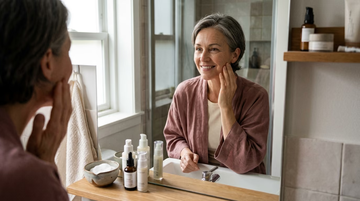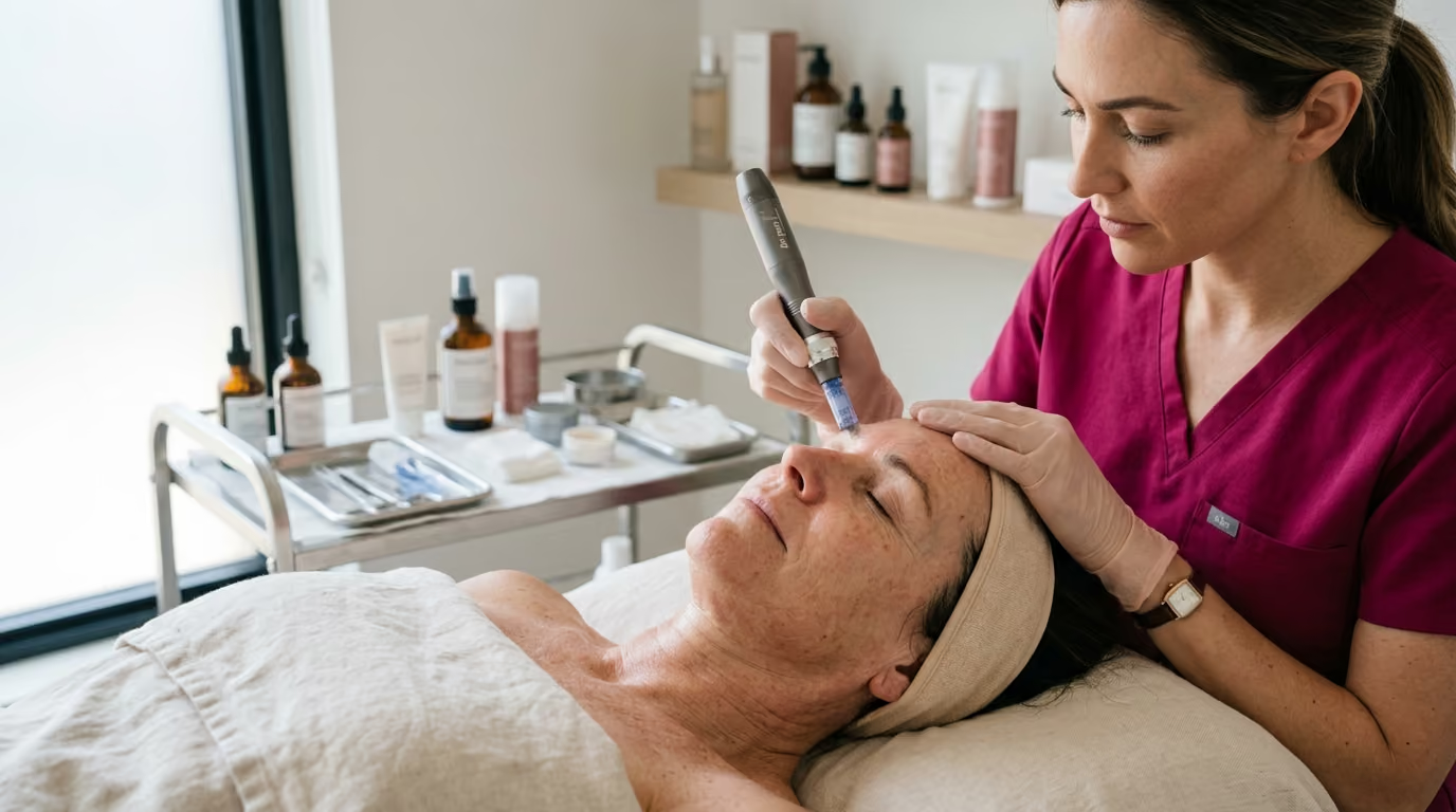

Three biological wound-healing phases drive collagen induction therapy to improve dermal thickness, smooth fine lines, and remodel aging skin effectively.





If you have ever searched whether microneedling can rebuild lost skin elasticity or if rolling tiny needles across your face is safe, you have likely encountered conflicting advice. Some sources promise dramatic structural renewal after a single session. Other sources warn of chronic redness, infection, and skin barrier destruction.
The truth lies within the precise biology of cellular wound healing. In this definitive guide, we review the scientific mechanisms, clinical evidence, device variations, and practical safety protocols of collagen induction therapy to help you make informed choices for your skin.
Clinical research categorizes microneedling as a minimally invasive dermatologic procedure that uses precise, sterile needles to trigger structural remodeling. The core scientific findings include:
Understanding how microneedling alters skin architecture requires looking below the surface. Human skin consists of two primary functional layers. The epidermis serves as the external barrier against environmental pathogens and moisture loss. The deeper dermis contains the extracellular matrix, which is primarily composed of type I and type III collagen, elastin fibers, and glycosaminoglycans. These structural proteins give young skin its mechanical strength and elasticity.
As chronological aging and sun exposure progress, fibroblast activity declines. Existing collagen fibers become fragmented, disorganized, and stiff. In our work reviewing beauty science and advanced optimization, we often find that procedures attempting to stimulate structural repair must strike a delicate balance. They must create enough stimulus to wake dormant fibroblasts without causing destructive scar tissue.
Microneedling achieves this through mechanical micro-trauma. When fine surgical needles puncture the skin, they do not slice tissue or burn it. Instead, they produce thousands of microscopic vertical channels that extend through the epidermis and into the upper dermis. This controlled physical trauma bypasses thermal damage entirely. The cellular response follows a predictable three-stage biological cascade.
The physical puncture immediately disrupts micro-capillaries, leading to localized platelet aggregation and micro-clot formation. Activated platelets release essential signaling proteins, including transforming growth factor-alpha, transforming growth factor-beta, and platelet-derived growth factor.
Neutrophils and monocytes migrate to the injury channels to clear cellular debris. These immune cells release additional cytokines that signal nearby fibroblasts to migrate toward the micro-injury site. Because the stratum corneum between puncture channels remains largely intact, the initial inflammatory phase resolves quickly. This rapid resolution minimizes prolonged swelling compared to traditional surgical dermabrasion.
Within several days of treatment, the proliferative phase begins. Fibroblasts become highly active, laying down a temporary matrix rich in type III collagen, fibronectin, and hyaluronic acid. Keratinocytes multiply and migrate across the microchannels, quickly restoring the physical barrier of the skin.
Simultaneously, the release of vascular endothelial growth factor stimulates angiogenesis. This formation of new, healthy micro-capillaries improves local nutrient and oxygen delivery to the newly forming tissue. The skin transitions from an emergency repair state into an active rebuilding state.
The final stage of healing continues for months following a single procedure. Over several weeks to months, matrix metalloproteinases break down the temporary type III collagen. Fibroblasts replace it with organized, mature type I collagen networks.
Elastin synthesis also increases, helping restore structural bounce and pliability to the dermal matrix. Because this maturation occurs slowly, visible improvements in dermal density and firmness develop over a period of three to six months. The biological goal is functional remodeling rather than acute swelling.
The mechanical action of microneedling creates thousands of temporary aqueous pathways through the stratum corneum. Under normal circumstances, large molecular weight ingredients cannot cross the lipid matrix of the skin barrier. The temporary microchannels significantly enhance the transdermal delivery of applied substances.
However, increased permeability introduces significant biological risk. When substances bypass the protective barrier and enter the vascular dermis, ingredients that are safe for surface application can trigger severe immune reactions. The safe application of topicals during microchannel patency requires pharmaceutical-grade purity and sterility.
Controlled clinical trials evaluating microneedling demonstrate consistent improvements across several facial aging markers. When we first started reviewing clinical trials on collagen supplementation and dermal procedures, our team was struck by how often commercial marketing misinterpreted the data. A study showing a minor increase in skin elasticity was suddenly headlined as a total age reset. It made us realize how desperately consumers need a clear translator for skin longevity and healthy aging research, someone who can explain precisely what a study proves and what it leaves unanswered.
A comprehensive review published in the Journal of Clinical and Aesthetic Dermatology evaluated the therapeutic breadth of microneedling across various clinical indications. The researchers confirmed measurable improvements in structural aging parameters, scar revisions, and overall tissue quality.
Dermatologic assessments using standardized aesthetic scales demonstrate that a series of three to six microneedling sessions can improve mild-to-moderate rhytides. Skin biopsies taken before and after treatment series show noticeable increases in dermal thickness and collagen fiber density.
Histological staining reveals thicker, more organized collagen bundles running parallel to the dermal-epidermal junction. These structural gains help soften fine lines around the periorbital and perioral regions. However, microneedling cannot reposition deep fat pads or resolve severe skin laxity, which require surgical intervention.
Skin texture irregularities and enlarged pore appearance often stem from structural laxity around the follicular infundibulum. When dermal collagen diminishes, the tissue surrounding the pore loses tension, causing the opening to appear larger.
By stimulating new collagen synthesis in the upper papillary dermis, microneedling tightens the supportive sheath surrounding the follicle. Clinical imaging reveals smoother surface topography and an optical reduction in visible pore diameter. These outcomes correlate with high patient satisfaction scores on the Global Aesthetic Improvement Scale.
Sun-damaged skin exhibits solar elastosis, where degraded elastin and irregular melanin distribution create a mottled appearance. Microneedling aids photodamaged skin through dual pathways. First, the rapid turnover of keratinocytes during the re-epithelialization phase encourages the shedding of superficial, hyperpigmented cells.
Second, the structural remodeling normalizes cell signaling between keratinocytes and melanocytes. Because the epidermis is not thermally ablated, microneedling carries a substantially lower risk of triggering rebound post-inflammatory hyperpigmentation compared to medium-depth chemical peels or resurfacing lasers. This makes it an appealing option for individuals with darker skin phototypes.
The medical utility of microneedling is thoroughly documented in the management of atrophic acne scars and surgical scars. Depressed scars form when abnormal fibrous bands pull the epidermis downward during poorly regulated inflammatory healing.
The physical insertion of needles mechanically disrupts these rigid fibrous strands. The subsequent release of growth factors encourages the deposition of new, organized collagen to elevate the scar base. Comparative studies show that for superficial rolling and boxcar scars, microneedling can yield improvements comparable to non-ablative fractional lasers with significantly shorter patient recovery times.
To understand how professional treatments fit into broader wellness, you can explore our resources on lifestyle, recovery, and environmental aging.
The clinical outcomes and safety profiles of collagen induction therapy vary based on the device selected. Practitioners utilize three primary categories of equipment, each defined by distinct operating mechanics and biological impacts.
Manual rollers are handheld cylindrical drums studded with hundreds of miniature surgical steel or titanium needles. As the operator rolls the drum across the face, the needles pierce the tissue. While manual rollers are inexpensive, they possess mechanical limitations.
Because the drum rotates, needles enter the skin at an angle and exit at an opposite angle. This curved path can create a micro-tearing or shear effect in the tissue, especially if the user applies excessive downward force. Furthermore, needle depth cannot be adjusted during the session, making it difficult to safely treat delicate areas like the lower eyelids or bridge of the nose.
Automated pens represent the clinical standard for mechanical collagen induction therapy. These devices utilize a high-speed reciprocating electric motor to drive a disposable cartridge of sterile needles vertically into the skin.
Because the needles enter and exit at an exact 90-degree angle, tissue shear is avoided, reducing surface micro-trauma and bruising. Automated pens allow the clinician to adjust the needle penetration depth in real time, ranging from 0.25 millimeters for thin periorbital tissue up to 2.5 millimeters for thick, scarred cheek tissue. The high frequency of the motor also minimizes procedural discomfort.
Radiofrequency microneedling combines physical needle puncture with the localized emission of thermal radiofrequency energy. Insulated or non-insulated needles penetrate the dermis, acting as electro-conductive conduits that heat the surrounding tissue to roughly 60 to 70 degrees Celsius.
This localized thermal coagulation causes immediate contraction of collagen fibers and triggers a robust structural healing response. While highly effective for skin tightening, radiofrequency microneedling is a sophisticated medical procedure with a distinct safety profile. The added heat alters tissue structure deeply, requiring specialized practitioner credentials to prevent complications.
While the scientific literature supports microneedling for skin longevity, objective medical evaluation requires highlighting what the research does not support.
First, clinical studies on microneedling frequently feature small sample sizes and variable methodologies. Researchers often use different needle lengths, varying session frequencies, and differing subjective scoring systems. This lack of standardization makes direct statistical comparisons between trials challenging.
Second, the durability of newly synthesized collagen remains subject to normal biological degradation. Microneedling does not halt intrinsic cellular aging or prevent future UV damage. Without ongoing sun protection and maintenance treatments every six to twelve months, structural gains will gradually diminish over time.
Third, significant controversy remains regarding topical add-on products. Many commercial clinics market the application of multi-ingredient peptide cocktails, botanical extracts, or unproven serums directly over newly created microchannels. Current clinical evidence supporting the additive value of these proprietary serums over simple saline or pharmaceutical hyaluronic acid remains limited.
More importantly, introducing non-sterile or unapproved cosmetic formulations into the deep dermis carries severe medical risks. Well-designed, large-scale clinical trials are still needed to determine optimal session spacing and standardized depth protocols for specific skin conditions.
A safe and effective microneedling protocol begins long before any needle touches the skin. Systematic planning minimizes complications and ensures the skin barrier heals quickly.
A medical professional must perform a comprehensive evaluation of the patient's dermatologic history, current medications, and scar tendencies. The American Academy of Dermatology highlights several absolute and relative contraindications:
Needle depth must be tailored to specific anatomical facial zones. The epidermis and dermis vary in thickness across the face:
Treatment sessions should be spaced four to six weeks apart. This interval ensures the inflammatory and proliferative phases conclude before applying a secondary injury. Rushing treatments compromises fibroblast viability and can lead to chronic, low-grade tissue inflammation.
Aftercare focuses on barrier protection, infection prevention, and UV avoidance. In the first 24 hours post-procedure, microchannels remain partially open:
Patients must be taught to monitor for clinical complications. Expected reactions include mild sunburn sensations, localized swelling, and transient pinpoint bleeding that resolve within 48 to 72 hours.
If you notice spreading erythema, localized warmth, increasing pain, or yellow crusting, consult your clinician promptly. These symptoms indicate potential bacterial infection requiring medical management.
To read more on practical skincare formulation, review our guides on nourishing the skin barrier from within.
The widespread availability of inexpensive dermal rollers and electric pens on e-commerce platforms has led to an increase in home microneedling. However, medical organizations, including the U.S. Food and Drug Administration and the American Academy of Dermatology, draw a clear distinction between clinical collagen induction and at-home rolling.
Consumer devices marketed for home use feature very short needle lengths, typically 0.1 to 0.25 millimeters. These superficial lengths only reach the outermost dead stratum corneum cells. They do not penetrate the vascular papillary dermis where fibroblasts reside.
As a result, home rolling cannot trigger the wound-healing cascade required for meaningful collagen synthesis. While it may temporarily enhance the absorption of surface hydrators, it will not remodel wrinkles or structural scars.
The primary danger of at-home microneedling is inadequate sanitation. In a clinical facility, automated pen tips are single-use, gamma-sterilized cartridges disposed of in biohazard containers immediately after treatment.
At-home rollers are reused multiple times. Household methods, like soaking a roller in isopropyl alcohol, do not achieve clinical-grade sterilization. Microscopic debris, sebum, and bacterial strains like Staphylococcus aureus accumulate between the needle bases.
Rolling these contaminated needles across the face can drive pathogens deep into the skin. This can lead to widespread folliculitis, impetigo, and the spread of viral conditions like flat warts or Herpes Simplex.
Consumer use often lacks standardized technique. Pressing too hard with a manual roller or dragging an electric pen across the skin can create irregular micro-tears. Repeated mechanical trauma over the same facial zones can cause linear "tram-track" scarring and localized hyperpigmentation.
Furthermore, applying conventional, non-sterile cosmetic serums immediately before or after home rolling introduces preservatives, emulsifiers, fragrances, and colorants directly into microchannels. The immune system identifies these foreign chemicals as invaders, which can trigger chronic foreign-body granulomas that require medical treatment to resolve.
Separating scientific facts from promotional marketing claims is essential when evaluating skin longevity procedures.
Reality: Indiscriminate depth increases complications without improving collagen synthesis. Penetrating into deep subcutaneous fat can damage facial nerves and microvascular networks without offering aesthetic benefit. Dermal remodeling occurs within the papillary and reticular dermis, which rarely requires exceeding 1.5 to 2.0 millimeters on the face. Proper clinical technique focuses on precision rather than aggressive depth.
Reality: Skincare formulations designed for surface application should never be driven into puncture channels. Standard cosmetic products contain preservatives like phenoxyethanol, penetration enhancers, thickeners, and fragrance compounds. When introduced into the vascular dermis, these ingredients can provoke allergic contact dermatitis, severe systemic sensitization, and granulomatous tissue reactions. Only pharmaceutical-grade, sterile saline or sterile, preservative-free hyaluronic acid solutions should be used.
Reality: Radiofrequency microneedling is an energy-based medical procedure, not a minor variation of mechanical needling. The addition of electro-thermal energy introduces risks of thermal burns, permanent fat atrophy, and subcutaneous scarring if administered with incorrect energy settings. The FDA explicitly categorizes radiofrequency microneedling as an advanced medical procedure that must never be attempted outside a qualified medical practice.
Reality: The fullness visible immediately following a microneedling session is transient inflammatory edema, not new collagen. True neocollagenesis and tissue remodeling are slow, complex biological processes. The synthesis, organization, and cross-linking of mature type I collagen fibers take several months to develop.
Initial swelling and redness generally subside within 48 to 72 hours, leaving the skin looking temporarily plump. True cellular repair begins around the second week, as fibroblasts lay down new extracellular matrix proteins. Visible structural improvements in fine lines, skin texture, and scar depth typically become noticeable between eight and twelve weeks. Collagen remodeling continues for up to six months after your final treatment session.
For general skin rejuvenation, mild photodamage, and subtle texture concerns, most clinical protocols recommend an initial series of three to four sessions spaced four to six weeks apart. For deeper atrophic acne scarring, surgical scars, or advanced photoaging, five to six sessions may be necessary. After completing the initial treatment series, maintenance sessions can be scheduled every six to twelve months to support ongoing skin longevity.
Yes. The mechanical stimulation and localized inflammatory signaling of microneedling can reactivate latent Herpes Simplex Virus residing in facial nerve endings. If you have a personal history of cold sores, inform your clinician prior to your appointment. They can prescribe a prophylactic course of oral antiviral medication to take before and after the procedure.
Mechanical microneedling is generally safer for Fitzpatrick skin types IV through VI than ablative lasers or deep chemical peels. Because the procedure does not use heat, it carries a lower risk of damaging melanocytes or triggering rebound post-inflammatory hyperpigmentation. However, conservative needle depths, gentle technique, and strict daily sun protection remain essential for minimizing pigment complications.
Potent active ingredients, including prescription retinoids, L-ascorbic acid, glycolic acid, salicylic acid, and mechanical scrubs, should be paused for five to seven days post-treatment. Resuming these ingredients too quickly can irritate the vulnerable re-epithelialized barrier and lead to contact dermatitis. Reintroduce one active product at a time once all redness, flaking, and sensitivity have resolved.
To expand your understanding of long-term skin health, explore our library of in-depth research resources.
If you are considering collagen induction therapy for your skin longevity plan, use this checklist to guide your decisions over the coming week:
Stay connected for research and practical guidance on skin, hair, collagen, nutrition and beauty longevity. Clear ideas for people who want to understand how appearance changes with age and make better-informed choices over time.


Understand your skin, hair and body better without chasing every new trend, treatment or promise.
explore the Blog