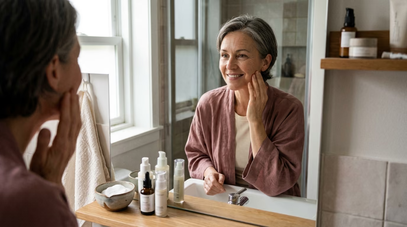

Many women expect simple dry skin during menopause, but falling estrogen levels fundamentally alter structural collagen, healing speed, and dermal elasticity.





Menopause is not a sudden skin crisis, nor is it a simple cosmetic issue that can be solved with a single cream. Clinically, menopause is defined as twelve consecutive months without menstruation, signaling the cessation of ovarian follicular activity. Perimenopause represents the transitional window before this milestone, marked by unpredictable hormonal fluctuations rather than a steady decline.
Hormonal skin aging is distinct from chronological aging and ultraviolet radiation damage, yet it interacts with both. Chronological aging alters cellular turnover across decades. Sun exposure breaks down structural fibers through oxidative stress. Menopause introduces a sharp reduction in systemic estrogen, altering the biological signals that maintain the skin matrix.
Understanding these biological shifts allows you to separate commercial marketing claims from physiological reality. This comprehensive guide examines how declining ovarian hormones alter dermal structure, surface hydration, skin sensitivity, and tissue repair. It provides a grounded, research-backed framework for managing skin health with realistic expectations.
Scientific investigations over the past several decades have clarified how hormonal shifts influence cutaneous biology. The primary findings from dermatological and endocrinological literature include:
Collagen provides the structural scaffolding for the entire dermis. Type I and Type III collagen fibers are responsible for tensile strength, firmness, and the physical thickness of the skin. Fibroblasts within the dermal layer rely on continuous molecular signals to manufacture these structural proteins and maintain tissue volume.
Estrogen acts as a primary signaling molecule for dermal fibroblasts. When estrogen binds to its receptors in the skin, it stimulates fibroblast proliferation and activates gene expression for collagen production. Furthermore, estrogen promotes the activity of tissue inhibitors of matrix metalloproteinases. These inhibitors prevent matrix metalloproteinases from breaking down healthy structural fibers prematurely.
When circulating estrogen levels drop during perimenopause and menopause, this synthetic signaling pathway weakens. Fibroblasts produce significantly less new Type I and Type III collagen. At the same time, the regulatory balance shifts, allowing matrix metalloproteinase enzymes to degrade existing structural fibers more rapidly.
The resulting net loss of collagen causes noticeable changes in tissue behavior. The dermis loses physical thickness and mechanical density. Fine lines become more pronounced because the underlying structural support has diminished. Furthermore, the skin takes longer to bounce back when pinched or compressed. Clinical assessments show that a skinfold on the back of the hand can take three to four times longer to return to its flat state in postmenopausal women compared to premenopausal women.
This structural decline does not occur evenly across the entire body. Areas that have accumulated chronic ultraviolet exposure, such as the face, neck, and forearms, experience the most noticeable changes. In these zones, hormonal collagen reduction combines with sun-induced matrix damage, creating accelerated structural slackness. You can read more about structural integrity within our collagen educational resources.
Skin hydration operates across two distinct biological levels: the deep dermis and the outer stratum corneum. Both levels depend heavily on hormonal support to maintain their water content and protective functions.
In the deeper dermis, hydration is governed by glycosaminoglycans, with hyaluronic acid serving as the primary water-binding molecule. These long sugar chains create a gel-like environment that holds water within the extracellular space, maintaining tissue turgor and fullness. As estrogen levels decline, dermal glycosaminoglycan production drops. The dermis loses its internal reservoir, causing the tissue to appear deflated and crêpey.
In the outer epidermis, the skin barrier relies on organized layers of dead skin cells embedded in a precise lipid matrix of ceramides, cholesterol, and free fatty acids. This structure prevents excessive transepidermal water loss and blocks environmental irritants. Estrogen supports the production of these essential surface lipids and encourages healthy cellular turnover.
When estrogen signaling falls, epidermal lipid synthesis decreases. The intercellular matrix becomes less organized, allowing vital moisture to escape into the environment at higher rates. The skin surface becomes rough, tight, and prone to flaking, particularly after washing with warm water or exposure to dry indoor air.
At the same time, elastic fibers within the dermis undergo progressive degeneration. While collagen provides tensile strength, elastin provides recoil, allowing the skin to snap back after stretching. With the loss of estrogen, elastic fibers become disorganized, fragmented, and stiffened by abnormal cross-linking.
The combined loss of dermal water, surface lipids, and functional elastin changes the physical characteristics of the skin. Laxity develops as structural support declines, while loss of elasticity prevents the skin from snapping back into position. Persistent dehydration deepens surface folds, making superficial wrinkles appear much more severe than they would in well-hydrated skin. Understanding these barrier mechanics is central to maintaining healthy skin longevity over time.
Many women observe that their skin becomes unpredictable and easily irritated during the menopausal transition. Skincare products that were tolerated for decades may suddenly cause burning, stinging, or redness. This heightened reactivity is rooted in architectural and immunological changes within the skin layers.
The dermal-epidermal junction is the undulating interface that anchors the outer epidermis to the supportive dermis beneath. In youthful skin, finger-like projections called rete ridges create a broad surface area for nutrient exchange and mechanical adhesion. As estrogen declines, this junction flattens, reducing the mechanical connection between the two layers.
In addition, research indicates that estrogen deficiency is associated with reduced expression of ezrin, a protein involved in maintaining cellular connections. With fewer cellular bridges and a flatter base, the epidermis becomes mechanically fragile. Minor friction, clothing tags, or light scratching can cause micro-tears and superficial abrasions that would not have occurred previously.
This structural vulnerability is compounded by changes in wound healing dynamics. Estrogen plays a direct role in every phase of tissue repair, including:
When estrogen is deficient, re-epithelialization slows down significantly. Minor cuts, blemishes, or procedure-induced wounds take longer to close and remain inflamed for extended periods. This delayed resolution increases the risk of post-inflammatory marks and persistent redness.
Skin sensitivity during menopause also stems from the compromised epidermal barrier. When the lipid layer is depleted, topical ingredients penetrate more deeply and unpredictably. Preservatives, fragrance compounds, and potent active ingredients reach sensory nerve endings in higher concentrations, triggering stinging sensations and neurogenic inflammation.
Changes in skin tone and pigment distribution are common complaints during the menopausal transition. Women often develop patchy discoloration, dark spots, or a dull, uneven complexion. However, these pigmentary changes rarely have a single cause.
Estrogen exerts complex regulatory effects on melanocytes, the specialized cells responsible for producing melanin pigment. Estrogen receptors are present on melanocytes, where they influence cellular activity and pigment synthesis. During perimenopause, erratic hormonal surges can stimulate localized melanocyte hyperactivity, contributing to melasma-like patches on the forehead, upper lip, and cheeks.
As estrogen permanently declines in postmenopause, overall baseline melanocyte density and activity tend to decrease. This can lead to a paler, less uniform complexion. However, localized clusters of damaged melanocytes often continue to produce pigment aberrantly, particularly in areas with extensive sun history.
Sun-induced lesions, known clinically as solar lentigines or age spots, are driven primarily by cumulative ultraviolet radiation exposure rather than hormones alone. Ultraviolet light damages the DNA of baseline melanocytes, causing them to permanently overproduce pigment in discrete patches. When hormonal thinning reduces epidermal thickness, these underlying pigment clusters become far more visible on the skin surface.
The alteration of the estrogen-to-androgen ratio during menopause also influences pigment and skin tone. While ovarian estrogen production drops dramatically, adrenal and ovarian androgen production declines much more gradually. This relative increase in androgenic signaling can alter oil production in the central face, occasionally triggering adult acne. Post-inflammatory hyperpigmentation then forms in the wake of these breakouts, lingering for months due to slower cellular turnover.
It is critical to distinguish benign pigment changes from serious dermatological conditions. Any pigmented lesion that is asymmetric, exhibits irregular borders, displays multiple colors, or changes rapidly must be evaluated by a dermatologist. Comprehensive skin health guidance always prioritizes clinical assessment over cosmetic self-treatment when assessing evolving spots.
The relationship between systemic menopausal hormone therapy and skin quality has been investigated for decades. Early observational studies suggested that women taking systemic estrogen had thicker skin, fewer wrinkles, and better dermal hydration than untreated peers. However, these early studies were often limited by small sample sizes, lack of control groups, and substantial selection bias.
The Kronos Early Estrogen Prevention Study, known as the KEEPS trial, provided rigorous randomized data regarding this question. An ancillary study within KEEPS evaluated 116 postmenopausal women who were within three years of their final menstrual period. Participants were randomized to receive oral conjugated equine estrogens, transdermal 17-beta estradiol, or a placebo, alongside cyclical progesterone when necessary.
The researchers tracked skin outcomes across four years of continuous treatment. The findings challenged several assumptions:
These findings do not mean that estrogen has no effect on skin biology. The KEEPS trial may have been underpowered to detect subtle cosmetic changes, or the four-year duration may have been too short to demonstrate visible separation in wrinkle depth. Furthermore, contemporary hormone regimens use lower, safer doses of estrogen compared to the high doses used in decades past, which may limit visible cutaneous effects.
Current guidelines from the North American Menopause Society clarify the appropriate role of systemic hormone therapy. Systemic treatment is indicated for the management of bothersome vasomotor symptoms, sleep disruption, and the prevention of bone loss in appropriate candidates. It is generally considered safe for healthy, symptomatic women under age 60 or within ten years of menopause onset.
However, professional medical guidelines do not recommend initiating systemic hormone therapy solely for cosmetic skin improvements. The systemic risks, including cardiovascular and thromboembolic considerations, outweigh the aesthetic benefits when no systemic symptoms exist. For women with an intact uterus, systemic estrogen must always be paired with a progestogen to protect the uterine lining from endometrial hyperplasia.
Because systemic hormone therapy carries systemic considerations, interest has shifted toward topical formulations that act locally on skin tissue. These include prescription topical steroid hormones, plant-derived phytoestrogens, and selective estrogen receptor modulators.
Topical estrogens, such as estriol or estradiol creams, can penetrate the stratum corneum and bind directly to cutaneous receptors. Small clinical trials indicate that topical application can improve dermal hydration, increase collagen content, and improve skin elasticity over several months.
However, topical hormones are not purely local treatments. Estrogen applied to the face or neck can be absorbed into the systemic bloodstream, especially when applied to thin, compromised skin barriers. The exact degree of systemic absorption from daily cosmetic facial use has not been definitively quantified in large trials. Consequently, topical estrogen remains a medical prescription requiring physician oversight, particularly for individuals with personal histories of hormone-sensitive conditions.
Phytoestrogens represent a non-steroidal alternative derived from botanical sources, including soy isoflavones, genistein, and daidzein. These compounds possess chemical structures that permit weak binding to human estrogen receptors, showing a distinct preference for estrogen receptor beta. Estrogen receptor beta is widely expressed in cutaneous fibroblasts and keratinocytes.
Clinical studies evaluating specific phytoestrogen derivatives have yielded modest but interesting findings:
While these findings are encouraging, they must be interpreted within their scientific context. These cosmeceutical studies relied heavily on subjective participant questionnaires rather than high-resolution histological biopsies. The trials had small sample sizes and lacked long-term safety data. Plant-derived compounds should be viewed as gentle supportive ingredients rather than direct equivalents to prescription hormone therapies. Exploring evidence-based lifestyle strategies provides a more holistic foundation for aging well.
Navigating skincare science requires a clear understanding of what current research can and cannot prove. Much of the popular discourse regarding menopausal skin oversimplifies complex physiological data.
A primary limitation in the existing literature is the difficulty of isolating hormonal aging from chronological and environmental factors. Chronological cellular senescence, cumulative solar radiation, air pollution, and dietary habits all occur concurrently with the menopausal transition. When a study evaluates postmenopausal skin, attributing structural changes exclusively to estrogen loss without controlling for forty years of sun exposure leads to skewed conclusions.
Additionally, demographic representation in historical dermatological trials has been narrow. Many fundamental studies on skin thickness and hormone therapy were conducted primarily on Caucasian women. As demonstrated by the KEEPS trial, baseline melanin levels, structural dermal density, and genetic background significantly influence how skin ages over time. Research findings from one demographic cohort cannot be universally applied to all skin types.
Many commercial cosmeceutical trials also suffer from methodological flaws:
Finally, researchers have not established universal, standardized values for changes in transepidermal water loss or wound healing rates specifically caused by menopause. While the biological mechanisms are verified, individual variation remains vast. Marketing claims that promise precise percentage increases in skin elasticity based on menopausal formulations should be met with careful skepticism.
An effective skincare strategy during perimenopause and postmenopause should prioritize barrier preservation, structural defense, and tissue comfort over aggressive exfoliation or trend-focused treatments.
The daily routine should be structured around four evidence-based pillars:
When considering clinical procedures, such as chemical peels, laser resurfacing, radiofrequency, or microneedling, careful planning is necessary. Because menopausal skin possesses a thinner epidermis, a flattened dermal-epidermal junction, and slower re-epithelialization, aggressive treatments carry higher risks of prolonged erythema or scarring.
Discuss conservative treatment parameters with your clinician. A series of low-intensity, staged procedures is generally safer and more effective than a single aggressive ablative treatment. Ensure your skin barrier is fully intact and hydrated for several weeks before undergoing any energy-based or needle-based clinical procedure.
Cutaneous health is closely connected to systemic physiological function. Broader lifestyle habits provide the essential raw materials for cellular maintenance:
Pervasive marketing narratives frequently distort the true biology of menopausal skin. Clarifying these misconceptions helps you make informed choices about your routine.
Reality: The magnitude of skin change varies substantially based on genetics, baseline pigmentation, lifetime ultraviolet exposure, smoking history, and local estrogen production in adipose tissue. While the biological mechanism is universal, the visible result is highly individualized.
Reality: Sensation is not an indicator of efficacy. Stinging and burning indicate that the epidermal barrier is compromised, allowing ingredients to trigger sensory nerve fibers. Chronic low-grade irritation degrades collagen and accelerates tissue sensitivity.
Reality: Well-designed clinical trials, including the KEEPS study, demonstrate that systemic hormone therapy does not reliably erase established wrinkles or prevent facial rigidity changes. Hormone therapy supports overall physiological health, but photoprotection and topical care remain essential for facial skin.
Reality: Phytoestrogens are biologically active compounds that bind human estrogen receptors. While they are generally gentle in cosmetic formulations, individuals with hormone-sensitive medical histories should still discuss their use with an oncologist or dermatologist.
Reality: Menopause can also cause adult acne due to relative androgen dominance, persistent flushing due to vasomotor instability, or chronic facial itching. Dryness is common, but it is only one component of a wider spectrum of possible presentations.
Navigating skin changes during perimenopause and menopause is most successful when coordinated with qualified medical professionals. A dermatologist or gynecologist can help distinguish normal hormonal shifts from underlying medical conditions.
Prepare for your consultation by tracking your symptoms, their onset, and any factors that worsen your comfort. Consider discussing the following questions during your visit:
Perimenopause involves significant hormonal fluctuations where estrogen levels drop periodically while ovarian and adrenal androgen levels remain relatively stable. This relative androgen excess can stimulate sebum production inside individual hair follicles. When this oil mixes with dead skin cells from a dry, sluggishly shedding epidermal surface, pores become clogged, resulting in localized adult breakouts alongside surface dryness.
While systemic hydration is important for general physiology, drinking excess water cannot resolve menopausal skin dryness. Dermal water retention depends on glycosaminoglycans like hyaluronic acid holding water in the matrix, while surface hydration depends on a lipid barrier preventing evaporation. Once these structural components decline due to lower estrogen, water simply evaporates through the skin unless supported by topical barrier lipids.
A vasomotor hot flash typically produces a sudden sensation of intense heat, rapid flushing across the face, neck, and chest, and often concludes with sweating within several minutes. Rosacea causes persistent, sustained facial redness, visible broken capillaries, and sometimes small inflammatory bumps localized to the central face that linger for days or weeks. Because both conditions can coexist during menopause, a dermatological evaluation is necessary for accurate diagnosis.
Yes, crêpey skin on the arms, legs, and décolletage shares the same fundamental biological origin as facial aging. It results from a simultaneous loss of dermal collagen, fragmented elastic fibers, and a significant decrease in dermal glycosaminoglycans. Because body skin has fewer sebaceous glands than the face, it produces fewer protective surface lipids, making thinning and dehydration particularly visible on the extremities.
Stay connected for research and practical guidance on skin, hair, collagen, nutrition and beauty longevity. Clear ideas for people who want to understand how appearance changes with age and make better-informed choices over time.


Understand your skin, hair and body better without chasing every new trend, treatment or promise.
explore the Blog