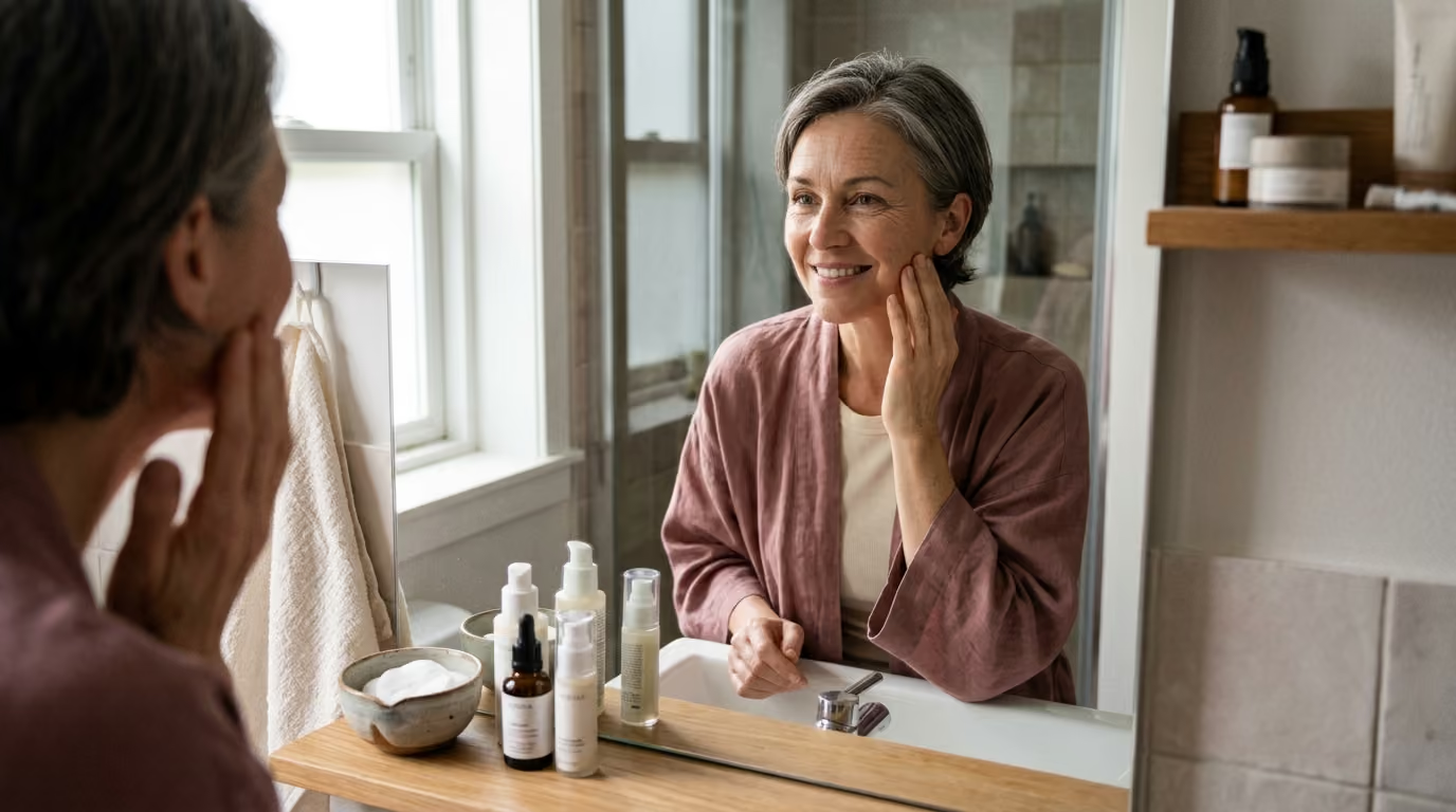

Targeted skin longevity routines become possible by distinguishing intrinsic chronological mechanisms from photoaging, environmental stressors.





Most people assume skin changes are driven either entirely by genetic heritage or entirely by sun exposure. This binary assumption leads to costly mistakes in skincare routines. When every line is treated as sun damage, people often overuse harsh resurfacing acids. When visible changes are dismissed as inevitable genetic decay, effective preventive habits are neglected.
Skin aging is not an all-or-nothing process. It is a layered biological reality where time-dependent physiological changes interact with cumulative environmental exposures. Understanding the distinct biological mechanisms of intrinsic aging and photoaging allows you to focus your energy on what you can realistically modify.
Skin aging represents the combined result of internal cellular timelines and external environmental damage. Chronological progression sets a baseline rate of tissue change across the entire body. External exposures then determine how early, how severely, and in what patterns visible changes appear.
Intrinsic aging is the gradual, time-dependent decline in physiological function that happens in all tissues. In sun-protected areas like the inner upper arm, skin undergoes structural shifts that remain largely invisible for decades. These changes stem from cellular senescence, telomere shortening, endogenous oxidative stress, and declining hormonal signals.
As skin cells age, their proliferative capacity drops. Basal keratinocytes in the epidermis divide more slowly, leading to a thinner epidermal layer. The dermo-epidermal junction flattens as rete ridges disappear, reducing the surface area available for nutrient exchange between the dermis and epidermis. This structural flattening makes mature skin more fragile and prone to mechanical shearing.
Within the dermis, fibroblast activity diminishes over time. Fibroblasts produce less type I and type III collagen, while endogenous matrix metalloproteinases continue their baseline breakdown. Unlike sun-damaged tissue, the remaining collagen and elastic fibers in intrinsically aged skin remain structurally organized, though they become sparse and thin. The microvascular supply also declines, which impairs thermoregulation and slows the clearance of metabolic waste.
Cellular senescence is a primary engine of intrinsic change. When cells reach their replicative limit, they enter a state of permanent growth arrest. These senescent cells secrete a mix of inflammatory cytokines, chemokines, and proteases known as the senescence-associated secretory phenotype. This chronic, low-grade inflammatory state degrades neighboring extracellular structures and impairs local stem cell renewal.
Photoaging is extrinsic aging accelerated by chronic exposure to solar ultraviolet radiation. Ultraviolet rays penetrate the skin at different depths, creating distinct biological cascades. Ultraviolet B radiation carries higher energy and penetrates the epidermis, directly damaging DNA by creating cyclobutane pyrimidine dimers. Ultraviolet A radiation penetrates deeply into the reticular dermis, generating massive amounts of reactive oxygen species.
When ultraviolet radiation strikes dermal fibroblasts and keratinocytes, it triggers cell-surface cytokine receptors. This activation stimulates intracellular signaling cascades, specifically mitogen-activated protein kinase pathways. These pathways activate transcription factor activator protein 1, which downregulates type I procollagen synthesis and upregulates matrix metalloproteinases.
Matrix metalloproteinases, including collagenase, gelatinase, and stromelysin, break down existing interstitial collagen fibers. Because the skin cannot repair this continuous enzymatic degradation completely, repeated solar exposure leads to fragmented, disorganized dermal scaffolding. This progressive loss of dermal structure is documented across our structural collagen changes research.
In addition to collagen degradation, photoaging produces a distinct histological phenomenon called solar elastosis. Instead of normal, organized elastic fibers, photoaged skin accumulates thickened, tangled, and morphologically abnormal elastin masses. These tangled fibers occupy spaces where healthy collagen once existed, resulting in coarse texture, deep furrows, and reduced mechanical recoil.
While ultraviolet radiation is the most studied external factor, it is not the only source of environmental damage. A comprehensive longevity strategy requires differentiating photoaging from other external exposures. Tobacco smoke, airborne particulate matter, and lifestyle disruptions each leave distinct biological signatures on the skin.
Tobacco smoke is an independent extrinsic risk factor that accelerates facial wrinkling. Cigarette smoke contains thousands of chemical compounds, including reactive hydrocarbons and heavy metals. When inhaled or deposited directly on tissue, these compounds trigger severe oxidative stress and systemic inflammation.
Nicotine causes acute cutaneous vasoconstriction, reducing microvascular blood flow to the epidermis and papillary dermis. This chronic reduction in capillary perfusion deprives fibroblasts of essential oxygen and micronutrients, impairing normal collagen synthesis. In addition, repetitive facial muscle contractions, such as pursing the lips during inhalation, create mechanical stress that deepens perioral lines.
Smoking also upregulates matrix-degrading enzymes while decreasing systemic antioxidant levels. Research confirms a dose-dependent relationship between lifetime pack-years and the severity of facial wrinkling. When combined with solar radiation, smoking acts synergistically to multiply tissue damage beyond the sum of either exposure alone.
Air pollution has emerged as a distinct contributor to extrinsic aging, particularly in dense urban environments. Particulate matter, polycyclic aromatic hydrocarbons, nitrogen dioxide, and ground-level ozone interact with the stratum corneum continuously. Ultrafine particles can penetrate hair follicles and compromise the cutaneous barrier.
Once inside the skin, these pollutants act via the aryl hydrocarbon receptor pathway. Activation of this receptor increases the production of reactive oxygen species, suppresses collagen synthesis, and triggers melanocyte hyperactivity. This biochemical cascade manifests clinically as mottled lentigines, uneven pigmentation, and impaired barrier hydration.
Unlike solar radiation, which causes deep textural furrows, pollution exposure is strongly associated with pigmentary changes. Studies evaluating urban populations show clear links between traffic-related soot and the formation of dark spots on the forehead and cheeks. Strategies to mitigate these urban exposures are detailed in our lifestyle and environmental recovery methods section.
Chronic sleep restriction and psychological stress alter skin physiology through neuroendocrine and immune mechanisms. Sleep is the primary biological period for cellular repair, growth hormone release, and metabolic clearance. When sleep duration is consistently inadequate, cutaneous recovery mechanisms become compromised.
Elevated psychological stress stimulates the hypothalamic-pituitary-adrenal axis, increasing circulating cortisol levels. Sustained cortisol elevations impair epidermal lipid synthesis, disrupt stratum corneum cohesion, and weaken the skin barrier. This biological state prolongs recovery from external irritants and exacerbates underlying inflammatory conditions.
Sleep deprivation also increases circulating inflammatory markers such as interleukin 6. While poor sleep may not directly cause deep structural wrinkles on its own, it reduces tissue resilience. This weakened resilience amplifies the damage caused by daytime ultraviolet and pollution exposures.
Determining whether your skin concerns stem primarily from intrinsic changes or external exposures requires a structured assessment. Because mature skin usually shows a mixed presentation, your goal is to identify which mechanism is dominant. Use the following four-step clinical assessment framework to evaluate your skin.
The most reliable clinical method for isolating external damage is comparing chronically sun-exposed areas with sun-protected skin. Useful comparisons include:
If your sun-protected inner arm shows smooth texture, uniform coloration, and only slight thinning, your facial lines are predominantly photo-induced. If both protected and exposed areas show uniform dryness, fragility, and loss of volume without deep furrows, intrinsic aging is the primary driver.
Examine the specific visual and tactile qualities of your skin to match them with their underlying biology.
Accurately calculating your cumulative exposure history helps explain visible discrepancies between individuals of identical chronological age. Review these historical factors:
Many people confuse temporary barrier impairment with accelerated biological aging. When the stratum corneum is stripped of natural ceramides and fatty acids, it loses water rapidly. This transepidermal water loss creates superficial roughness, flaking, and exaggerated fine dehydration lines.
These superficial dehydration lines disappear once proper barrier lipid levels and hydration are restored. Structural wrinkles caused by dermal collagen loss, however, remain visible regardless of surface hydration. Always repair an irritated, compromised skin barrier before evaluating deeper tissue architecture.
Understanding what clinical studies have proven prevents reliance on marketing narratives. Rigorous evidence demonstrates clear parameters for what interventions can and cannot achieve.
The strongest clinical evidence for preventing extrinsic aging comes from a randomized controlled trial conducted by Green and colleagues in Queensland, Australia. This four-and-a-half-year study followed 903 adult participants to evaluate whether daily broad-spectrum sunscreen application altered skin aging patterns compared to discretionary use.
Skin aging was objectively measured using microtopographic silicone impressions of the dorsal hands, graded on a validated scale. At the conclusion of the trial, the daily-use group showed 24% less skin aging progression than the discretionary-use control group. In fact, participants in the daily-use cohort showed no detectable increase in skin aging over the entire study window.
This trial provides definitive evidence that daily photoprotection prevents the accumulation of new dermal matrix damage. However, the data must be interpreted carefully. Daily sunscreen prevented additional aging progression, but it did not erase decades of established solar elastosis.
The epidemiological connection between air pollution and premature facial aging was established in the SALIA cohort study by Vierkötter and colleagues. This study evaluated 400 women aged 70 to 79 years to quantify the relationship between chronic traffic-related exposures and visible skin changes.
The investigators documented that an interquartile-range increase in soot and traffic particles was associated with 16% to 22% more pigment spots on the forehead and cheeks. Nitrogen dioxide exposure was similarly correlated with increased lentigines and more pronounced nasolabial folds. Subsequent analyses have reinforced that particulate pollution primarily drives pigmentary dyschromia rather than isolated deep wrinkling.
The biological relationship between sleep restriction and skin function was quantified in controlled laboratory models by Altemus and colleagues. Researchers evaluated cutaneous barrier recovery rates after tape stripping in healthy individuals subjected to sleep disruption compared to well-rested controls.
Participants experiencing sleep restriction required an average of 5.0 days to achieve full barrier recovery, compared to 4.2 days in the control group. The data demonstrated that sleep restriction significantly impairs acute barrier repair kinetics and increases systemic inflammatory cytokines. While sleep disruption does not directly break down collagen, it impairs the tissue's capacity to repair daily environmental damage.
Translating scientific papers into daily personal care requires acknowledging the boundaries of current research. Overinterpreting clinical studies leads to unrealistic expectations and misallocated resources.
Human skin aging studies face significant methodological limitations due to exposure confounding. In free-living populations, individuals exposed to high levels of ultraviolet light are frequently exposed to outdoor air pollution and temperature fluctuations. Similarly, cigarette smokers often share lifestyle, dietary, and sleep variables that differ from non-smokers.
Isolating the precise percentage of tissue aging caused by a single variable is virtually impossible outside of controlled animal models. While reviews frequently claim that ultraviolet light accounts for up to 80% of visible skin aging, this number varies depending on geographic latitude, occupational history, and genetic background. It should be understood as a general estimate rather than an exact biological law.
A major limitation of early dermatological research is its disproportionate focus on light-skinned individuals of European descent. Skin phenotypes age through different dominant morphological patterns depending on melanin content and baseline dermal thickness.
Research confirms that darker skin phenotypes possess higher baseline melanin density, providing natural photoprotection against ultraviolet-induced erythema and collagen breakdown. Consequently, skin of color is less susceptible to early coarse wrinkling and solar elastosis. However, it is far more prone to pigmentary dyschromia, melasma, and post-inflammatory hyperpigmentation. Longevity guidance must adapt to these biological variations rather than applying light-skin models universally.
Much of our mechanistic knowledge regarding cellular senescence and antioxidant pathways comes from in vitro cell cultures. Exposing isolated fibroblasts in a petri dish to concentrated ultraviolet radiation or botanical extracts does not mirror the complex biology of living human skin.
Topical compounds that neutralize free radicals in laboratory assays often fail to penetrate the stratum corneum in therapeutic amounts. Furthermore, the skin possesses robust endogenous antioxidant defense networks that behave differently than isolated topicals. Claims asserting that a specific topical antioxidant can neutralize all environmental damage should be viewed with healthy skepticism.
Achieving long-term skin health requires organizing interventions by their proven efficacy. Rather than adopting an unmanageable routine, structure your habits into a three-tiered prioritization framework.
The first tier contains non-negotiable interventions supported by the highest level of randomized clinical evidence. These habits prevent the primary drivers of extrinsic cellular degradation:
The second tier focuses on supporting endogenous repair mechanisms and cellular recovery. These lifestyle factors maintain the physiological environment necessary for tissue longevity.
Prioritize consistent, high-quality sleep by maintaining a regular sleep schedule and optimizing your sleep environment. Restorative sleep supports natural growth hormone pulses and normalizes immune function, ensuring efficient daily barrier recovery. Manage systemic stress through regular physical exercise and mindfulness practices to keep baseline cortisol levels in check.
Minimize your exposure to urban particulate pollution when possible. Avoid strenuous outdoor exercise adjacent to congested traffic corridors during peak rush hours. Within our broader evidence-based skincare research, maintaining an intact lipid barrier is the primary biological shield against environmental particle penetration.
During our investigation into environmental aging, our team tested how various lifestyle factors impact skin barrier recovery. In our experience, the data clearly show that simple habits like sleep and basic hydration often outperform the most expensive topical treatments. This reinforced our commitment to emphasizing foundational health over product hype.
The third tier introduces individualized topical agents and clinical interventions to address specific structural concerns.
Incorporate proven topical ingredients, such as prescription retinoids or over-the-counter retinol, to stimulate procollagen synthesis and normalize epidermal turnover. Use targeted, gentle ingredients like niacinamide or azelaic acid to regulate melanocyte activity if post-inflammatory hyperpigmentation or lentigines are present. For deeper structural loss, consult a board-certified dermatologist to discuss clinical modalities.
Most importantly, establish a routine of regular skin self-examinations. Promptly refer any changing, asymmetric, bleeding, or non-healing cutaneous lesions to a medical professional. Preserving skin longevity is fundamentally rooted in preserving tissue health and preventing malignancy.
Explore our collection of skin longevity and healthy aging resources for deeper guides on optimizing individual routines.
Misconceptions regarding skin aging waste time, money, and emotional energy. Separating clinical reality from beauty marketing claims helps you make grounded, confident choices.
Dehydration lines are superficial creases in the stratum corneum caused by temporary water loss. They typically appear alongside a tight, dull, or slightly flaky skin surface and resolve within hours of applying effective humectants and occlusives. Structural wrinkles from intrinsic aging or photoaging persist regardless of surface hydration levels because they stem from deeper dermal collagen loss and elastin degradation.
Ultraviolet B rays are largely blocked by clouds and standard window glass, reducing your risk of sunburn. However, up to 80% of ultraviolet A rays penetrate light cloud cover, and standard glass does not block longer UVA wavelengths. If you spend significant time near windows or outdoors on overcast days, broad-spectrum sunscreen remains an essential tool to prevent cumulative oxidative dermal damage.
Intrinsic aging is a continuous biological process that begins in early adulthood. Starting around your mid-twenties, dermal collagen production drops by roughly 1% per year, and epidermal turnover gradually slows. These subtle cellular changes take several decades to manifest as visible fine lines or skin thinning, which is why early preventive photoprotection is so valuable.
Facial exercises and massage cannot rebuild degraded collagen matrices, clear solar elastosis, or repair damaged cellular DNA. While massage may temporarily increase local microvascular blood flow, aggressive or repetitive facial contractions can actually deepen dynamic expression lines. Long-term skin longevity relies on cellular protection and matrix preservation rather than mechanical manipulation.
Dietary habits influence systemic oxidative stress, advanced glycation end-product formation, and baseline inflammation. High consumption of refined sugars promotes collagen cross-linking through glycation, which stiffens the dermal matrix. Conversely, a diet rich in essential fatty acids, dietary antioxidants, and adequate protein provides the building blocks necessary for continuous cellular repair and optimal barrier lipid synthesis.
Any new, growing, asymmetrical, or irregularly pigmented spot should be evaluated by a dermatologist or qualified healthcare professional. While many brown spots are benign solar lentigines or seborrheic keratoses, distinguishing benign pigmentation from early lentigo maligna or other cutaneous malignancies requires professional dermoscopic examination.
Navigating skin longevity requires shifting away from unrealistic promises and grounding your daily habits in verified biological realities. For further reading across our research archive, explore our science-led longevity articles.
Stay connected for research and practical guidance on skin, hair, collagen, nutrition and beauty longevity. Clear ideas for people who want to understand how appearance changes with age and make better-informed choices over time.


Understand your skin, hair and body better without chasing every new trend, treatment or promise.
explore the Blog