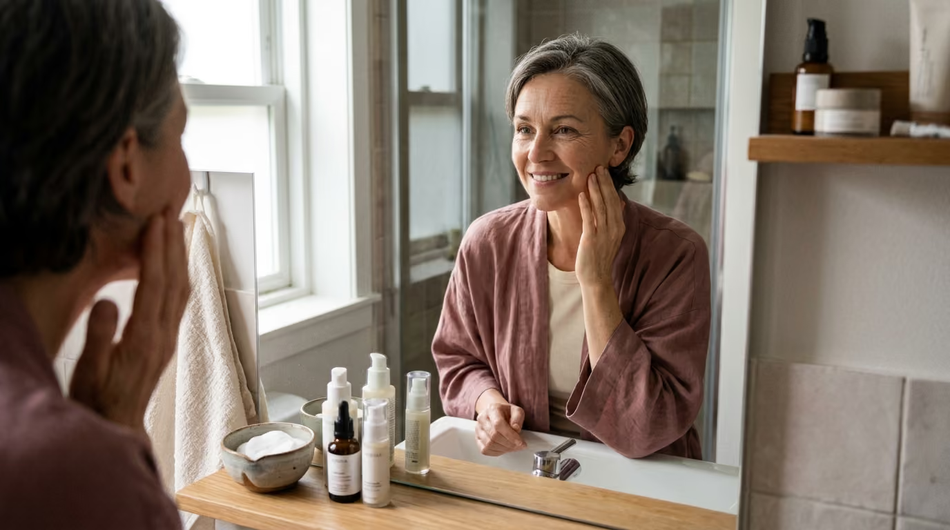

Dermal elasticity gradually diminishes as ultraviolet radiation and metabolic glycation break down the structural collagen matrix supporting healthy skin.





Most conversations about skin health treat collagen like a simple bank account. The prevailing assumption suggests that youthful skin has a large deposit of protein, aging drains the balance, and applying or swallowing collagen simply refills the account.
This view is biologically inaccurate. Skin aging is rarely just a raw shortage of collagen molecules. It is an architectural failure involving synthesis, enzymatic breakdown, structural cross-linking, and cellular signaling within the extracellular matrix.
Understanding how collagen functions requires examining the dermis as an active, living ecosystem. When you look at the mechanical tension of fibroblasts, the enzymatic activity of matrix metalloproteinases, and the molecular impact of ultraviolet light, the path to long-term skin health becomes clear. Preserving dermal integrity is far more achievable than attempting to replace lost tissue after structural collapse occurs.
Collagen serves as the primary structural protein of human connective tissue. In the skin, it forms the dense fibrous network that gives the dermis its tensile strength, physical resilience, and structural volume. Without an organized collagen network, the skin cannot resist mechanical shearing, retain deep hydration, or support overlying epidermal structures.
The dermis contains several distinct collagen variants, but types I and III dominate the cutaneous scaffold. Type I collagen accounts for roughly 80 to 85 percent of adult dermal collagen, providing rigid tensile support. Type III collagen makes up approximately 10 to 15 percent, offering elasticity and compliant structural support, particularly in vascular walls and newly healing wounds.
Dermal collagen does not exist in isolation. It resides inside an intricate extracellular matrix composed of elastin fibers, proteoglycans, glycosaminoglycans, and structural glycoproteins. This matrix acts as a functional biomechanical scaffold that physically anchors dermal fibroblasts, the specialized cells responsible for producing structural proteins.
The biological synthesis of collagen is a multi-step pathway requiring precise intracellular assembly and extracellular maturation:
The process begins in the fibroblast nucleus. Chemical signals, mechanical stretch, and systemic hormones stimulate the transcription of specific collagen genes, primarily COL1A1 and COL1A2. These genetic instructions are transcribed into messenger RNA and transported into the cytoplasm.
Ribosomes along the rough endoplasmic reticulum translate the RNA sequence into precursor protein chains known as pre-procollagen. These individual polypeptide chains contain repeating amino acid triplets, characteristically glycine followed by proline or hydroxyproline. Glycine, being the smallest amino acid, is positioned at every third residue to allow tight spatial packing.
Inside the endoplasmic reticulum, specific enzymes modify the newly formed polypeptide chains. Prolyl hydroxylase and lysyl hydroxylase add hydroxyl groups to proline and lysine residues. Ascorbic acid, commonly known as vitamin C, acts as an essential electron donor and cofactor for these hydroxylase enzymes. Without adequate vitamin C, this hydroxylation stalls, yielding unstable chains that cannot fold correctly.
Three hydroxylated procollagen peptide chains wind around one another to form a stable, right-handed triple helix. Hydrogen bonds formed between the hydroxylated amino acids hold this helical structure together. The resulting molecule is called procollagen, which possesses loose, non-helical peptide extensions on both ends to prevent premature assembly inside the cell.
The fibroblast packages the procollagen molecule into secretory vesicles and transports it across the cell membrane into the extracellular space. Once outside the cell, specific extracellular peptidase enzymes cleave the terminal propeptide caps from both ends of the molecule. This cleavage converts procollagen into insoluble tropocollagen.
Individual tropocollagen molecules spontaneously align in a staggered, overlapping pattern to form microfibrils, which group together into larger collagen fibrils. An extracellular enzyme called lysyl oxidase initiates covalent cross-linking between adjacent lysine and hydroxylysine residues. These enzymatic cross-links give the final collagen fiber its high tensile strength and physical durability.
This intricate assembly process highlights an important biological reality. Generating new structural support requires active cellular machinery, mechanical stability, and biochemical cofactors. It cannot be achieved simply by rubbing structural proteins on the epidermal surface. To explore more about these structural dynamics, read our guide on collagen and structural aging.
Collagen decline is not a sudden event that begins in midlife. It represents a continuous, lifelong shift in the balance between matrix production and matrix degradation.
From early adulthood, natural collagen synthesis in human skin decreases by roughly 1 to 1.5 percent per year. This gradual reduction stems from progressive changes in dermal fibroblast function. As fibroblasts age chronologically, their synthetic capacity slows, cellular replication declines, and their sensitivity to growth factor stimulation weakens.
A major biological shift occurs during the menopausal transition in women. Estrogen plays an active regulatory role in connective tissue maintenance by stimulating fibroblast proliferation, promoting procollagen gene transcription, and maintaining dermal hyaluronic acid content.
When circulating 17-beta-estradiol levels decline during perimenopause and menopause, dermal collagen loss accelerates significantly. Research published in historical dermatological literature indicates that skin collagen content can decline by roughly 2.1 percent per year following menopause. Studies have documented a cumulative loss of nearly 30 percent of dermal collagen during the initial five years post-menopause.
It is essential to differentiate intrinsic chronological aging from extrinsic photoaging. Intrinsic aging represents the biological clock operating in sun-protected skin, such as the inner upper arm. In intrinsically aged tissue, the collagen network becomes thinner and less dense, but the remaining fibers remain relatively straight and well-organized.
Extrinsic photoaging, caused primarily by cumulative ultraviolet radiation, produces a chaotic and structurally damaged matrix. Photoaged skin does not just suffer from reduced collagen volume. It exhibits extensive fiber fragmentation, abnormal protein aggregation, and the accumulation of non-functional elastic material known as solar elastosis.
Beyond simple quantity, the mechanical properties of dermal tissue depend entirely on collagen architecture. In youthful skin, collagen bundles are arranged in a dynamic, basket-weave orientation that flexes and recoils under physical stress. As collagen degrades, this architectural organization breaks down, leaving behind a fragmented matrix that cannot support the overlying epidermis.
This loss of structural continuity flattens the dermal-epidermal junction. The interlocked epidermal ridges and dermal papillae that facilitate nutrient transport between skin layers become smooth, diminishing mechanical stability. The visible result includes skin laxity, fine lines, hollowed contours, and a loss of elastic recoil. For a broader overview of how cutaneous structures change over time, see our resources on skin longevity and healthy aging.
Understanding the biology of collagen degradation is critical because preventing matrix loss is far more effective than attempting to rebuild collapsed tissue. Matrix degradation is driven by specific biochemical processes: ultraviolet radiation, cigarette smoke exposure, and metabolic glycation.
Solar ultraviolet radiation represents the single most destructive extrinsic factor affecting cutaneous connective tissue. Both UVA and UVB wavelengths drive dermal degradation through distinct but complementary mechanisms.
UVB rays possess shorter wavelengths and higher energy, penetrating the epidermis and upper papillary dermis where they directly damage cellular DNA. UVA rays possess longer wavelengths, penetrating deeply into the reticular dermis where structural collagen networks and fibroblasts reside.
When ultraviolet light strikes dermal fibroblasts and keratinocytes, it generates reactive oxygen species. These reactive molecules activate intracellular signaling pathways, specifically the AP-1 and NF-kB transcription factor complexes. Once activated, these transcription factors trigger the expression of matrix metalloproteinases, a family of zinc-dependent endopeptidases.
Matrix metalloproteinases degrade extracellular structural proteins. MMP-1, also known as interstitial collagenase, makes the initial cleaving cut across the triple helix of intact types I and III collagen fibers. Once cleaved by MMP-1, the unwound collagen fragments become vulnerable to further destruction by MMP-3 (stromelysin-1) and MMP-9 (gelatinase B).
Simultaneously, active AP-1 signaling blocks the transforming growth factor-beta pathway, the primary biochemical signal that instructs fibroblasts to produce new procollagen. Ultraviolet exposure creates a dual catastrophe: it accelerates the enzymatic destruction of existing collagen while shutting down the cellular machinery needed to replace it.
When collagen fibers are cleaved into fragments, fibroblasts lose physical attachment to their surrounding matrix. Dermal fibroblasts require mechanical tension to function properly; they must stretch between intact structural fibers to maintain procollagen synthesis. When the surrounding matrix collapses, fibroblasts collapse inward, rounding up and shifting their biological state from matrix synthesis to sustained enzyme secretion.
Tobacco smoke delivers a concentrated mixture of thousands of toxic compounds directly into systemic circulation. Nicotine causes acute peripheral vasoconstriction, significantly reducing cutaneous microvascular perfusion and oxygen delivery to the dermis. Dermal fibroblasts require continuous oxygen and nutrient delivery to sustain procollagen synthesis.
Furthermore, tobacco smoke introduces reactive free radicals that deplete endogenous cutaneous antioxidants such as ascorbic acid and alpha-tocopherol. Research examining advanced glycation end products in dermal tissue indicates that the tobacco metabolite nornicotine reacts directly with matrix proteins. This reaction accelerates the non-enzymatic modification of collagen, leading to premature structural stiffness, impaired wound healing, and accelerated skin wrinkling.
Metabolic health exerts a profound influence on dermal structural integrity through a biochemical process called glycation. Glycation occurs when circulating reducing sugars, such as glucose and fructose, react non-enzymatically with the amino groups of long-lived proteins.
Because dermal collagen has a slow turnover rate with a half-life measured in years, it is uniquely susceptible to this chemical modification. Over time, early glycation products undergo complex rearrangements, oxidation, and dehydration reactions to form permanent structures called advanced glycation end products, or AGEs.
These advanced glycation end products form abnormal, permanent cross-links between adjacent collagen fibrils. Unlike the flexible enzymatic cross-links created by lysyl oxidase, glycation cross-links make the collagen matrix excessively rigid, brittle, and unable to flex under biomechanical tension.
Glycated collagen resists natural enzymatic remodeling, meaning damaged structural proteins cannot be cleared and replaced by fibroblasts. Additionally, AGEs bind to specific cell-surface receptors called RAGE (receptors for advanced glycation end products) on fibroblasts and endothelial cells.
This binding activates inflammatory signaling cascades that promote continuous, low-grade MMP secretion and sustained oxidative stress. To learn more about the broader science of cellular aging, explore our articles on beauty science and cellular pathways.
The scientific literature contains substantial evidence regarding interventions designed to preserve, stimulate, or remodel dermal collagen. Differentiating between rigorous clinical outcomes and promotional marketing requires evaluating the data objectively.
Oral collagen supplementation remains one of the most widely debated topics in beauty longevity. The primary misconception assumes that ingested collagen travels directly from the digestive tract into the skin as whole fibers. In reality, the digestive system breaks ingested proteins down into single amino acids, dipeptides, and tripeptides.
Specific bioactive dipeptides, notably prolyl-hydroxyproline and hydroxyprolyl-glycine, are absorbed across the intestinal barrier and enter systemic circulation. Controlled pharmacokinetic studies demonstrate that these specific peptide fragments can reach cutaneous tissue.
Rather than serving solely as raw building materials, these circulating peptides act as biological signaling molecules. They bind to surface receptors on dermal fibroblasts, stimulating the cells to produce hyaluronic acid, elastin, and procollagen.
A 2019 systematic review published in the Journal of Drugs in Dermatology evaluated 11 randomized, placebo-controlled trials involving a total of 805 participants. Hydrolyzed collagen doses ranged from 2.5 grams to 10 grams daily for durations spanning 8 to 24 weeks. The researchers concluded that oral collagen supplementation supported statistically significant improvements in skin elasticity, hydration, and dermal collagen density.
A subsequent meta-analysis of 10 randomized controlled trials encompassing 646 participants confirmed significant gains in hydration and elasticity, identifying an average effective dose of roughly 3.5 to 4 grams per day.
While these findings confirm measurable biological activity, they must be interpreted realistically. Oral peptides provide modest, incremental improvements in elasticity and skin hydration. They do not rebuild deeply collapsed dermal folds or replicate the structural lifting of procedural interventions.
Topical retinoids, particularly all-trans retinoic acid (tretinoin), represent the most rigorously validated topical agents for collagen preservation and renewal. Retinoids bind to specific nuclear receptors in cutaneous cells, known as retinoic acid receptors and retinoid X receptors.
Once bound, retinoids directly regulate gene transcription. In the dermis, tretinoin stimulates fibroblasts to increase procollagen synthesis, specifically types I and III. Histological studies analyzing human punch biopsies confirm substantial increases in procollagen content after 3 to 12 months of consistent topical application.
Simultaneously, retinoids suppress the activation of AP-1, blocking the upregulation of MMP-1, MMP-3, and MMP-9 following ultraviolet exposure. This dual action stimulates new protein synthesis while protecting existing matrix architecture.
Energy-based devices and micro-injury modalities stimulate neocollagenesis by initiating a controlled wound-healing response:
Interpreting collagen research requires a clear understanding of study limitations and methodological constraints.
A significant limitation in the oral collagen literature involves commercial sponsorship. Many published trials are funded by supplement manufacturers or raw ingredient suppliers. While industry funding does not automatically invalidate findings, it introduces potential publication bias, where neutral or negative results may remain unpublished.
Furthermore, study methodologies vary considerably across the published literature:
Many clinical studies rely on surrogate instrumental endpoints, such as corneometer hydration readings, cutometer elasticity scores, or high-frequency ultrasound echo-density. These measurements provide useful objective data, but they do not always translate into dramatic, visible improvements in facial volume, skin laxity, or deep structural folds.
A significant gap exists between laboratory cell culture studies and living human skin. Demonstrating that a topical botanical extract or isolated peptide stimulates procollagen mRNA transcription in an in vitro Petri dish of isolated fibroblasts does not prove clinical efficacy. The molecule must cross an intact stratum corneum barrier, evade enzymatic degradation, penetrate into the deep vascular dermis, and reach target fibroblasts in an active biological concentration.
Finally, procedural studies often examine specific, limited demographics, predominantly lighter skin types (Fitzpatrick types I through III). Energy-based devices carry different risk profiles in deeper skin tones (Fitzpatrick types IV through VI), where thermal injury carries a higher risk of post-inflammatory hyperpigmentation or keloid scarring. Clinical outcomes must always be evaluated within the context of individual skin physiology.
A scientifically grounded approach to collagen preservation prioritizes structural defense, metabolic support, targeted topical signaling, and realistic nutritional foundations. To explore the broader foundations of cutaneous health, see our overview of evidence-based skin health.
Preventing ongoing structural breakdown is the foundation of any collagen preservation plan. No topical serum or procedural treatment can outpace the continuous matrix degradation caused by unprotected ultraviolet exposure.
Daily application of broad-spectrum sunscreen protecting against both UVA and UVB rays is non-negotiable. Select formulas that offer high UVA protection, verified by PA++++ ratings or high percentages of zinc oxide, because UVA is the primary driver of deep dermal MMP activation. Apply photoprotection every morning to all exposed areas, including the neck, chest, and hands.
To support structural renewal, incorporate active ingredients with validated clinical mechanisms:
Provide the systemic building blocks required for connective tissue maintenance:
Navigating the beauty industry requires separating physiological reality from marketing narratives.
The Reality: The molecular weight of an intact collagen molecule is roughly 300,000 Daltons. The human stratum corneum operates an effective transdermal penetration limit of approximately 500 Daltons.
Applying an intact collagen cream creates a hydrating, moisture-binding film on the surface of the stratum corneum, which temporarily softens the visual appearance of fine dehydration lines. However, these large molecules cannot penetrate the epidermal barrier to integrate into the dermal matrix.
The Reality: Ingested collagen is hydrolyzed by gastric acids and pancreatic proteases into free amino acids, dipeptides, and tripeptides. The body does not transport intact collagen protein directly from the gut into facial tissue.
Any physiological benefits from hydrolyzed collagen peptides stem from specific circulating bioactive fragments that act as signaling messengers, encouraging fibroblasts to produce new matrix proteins.
The Reality: Collagen quality and architectural organization matter far more than sheer quantity. Severely scarred tissue or heavily glycated dermis contains dense concentrations of collagen, but these fibers are abnormally cross-linked, stiff, and structurally brittle. Healthy skin requires an organized, pliable basket-weave architecture that flexes under mechanical stress.
The Reality: Visible tightening immediately following a laser or radiofrequency procedure reflects acute thermal contraction of preexisting collagen fibrils and transient tissue edema.
True neocollagenesis and matrix remodeling require active fibroblast transcription, protein secretion, and enzymatic cross-linking. This biological cascade develops gradually over a period of three to six months.
Topical products containing large intact collagen molecules cannot rebuild the dermal matrix because they cannot penetrate the stratum corneum.
However, specific signaling molecules, most notably prescription tretinoin and stabilized retinol, cross the epidermal barrier and bind to nuclear receptors in dermal fibroblasts. This binding stimulates direct procollagen gene transcription and suppresses collagen-degrading enzymes, promoting gradual structural renewal over months of consistent use.
Most randomized, placebo-controlled clinical trials document statistically significant improvements in skin hydration, elasticity, and dermal density after 8 to 12 weeks of daily supplementation.
Doses typically range from 2.5 grams to 5 grams of hydrolyzed collagen peptides or collagen tripeptides. Discontinuing supplementation generally results in a gradual return to baseline measurements over several months. To read more about oral supplementation research, visit our library of collagen science guides.
Vitamin C is an obligate cofactor for the prolyl and lysyl hydroxylase enzymes that stabilize the collagen triple helix. Severe vitamin C deficiency impairs collagen folding, leading to connective tissue breakdown.
However, once systemic plasma levels are saturated, consuming high-dose oral vitamin C supplements does not produce a proportional increase in dermal collagen synthesis in individuals who already consume an adequate, balanced diet.
Facial massage can transiently increase localized cutaneous blood flow, delivering oxygen and nutrients to tissues, while creating temporary mechanical stimulation.
However, robust clinical evidence demonstrating that manual massage or facial exercise produces long-term, structurally meaningful neocollagenesis remains limited. Excessive, aggressive mechanical pulling can stretch delicate cutaneous elastic fibers without stimulating organized collagen synthesis.
Protecting dermal collagen requires moving past simplistic marketing claims and working with the natural biological systems that govern connective tissue health.
Stay connected for research and practical guidance on skin, hair, collagen, nutrition and beauty longevity. Clear ideas for people who want to understand how appearance changes with age and make better-informed choices over time.


Understand your skin, hair and body better without chasing every new trend, treatment or promise.
explore the Blog