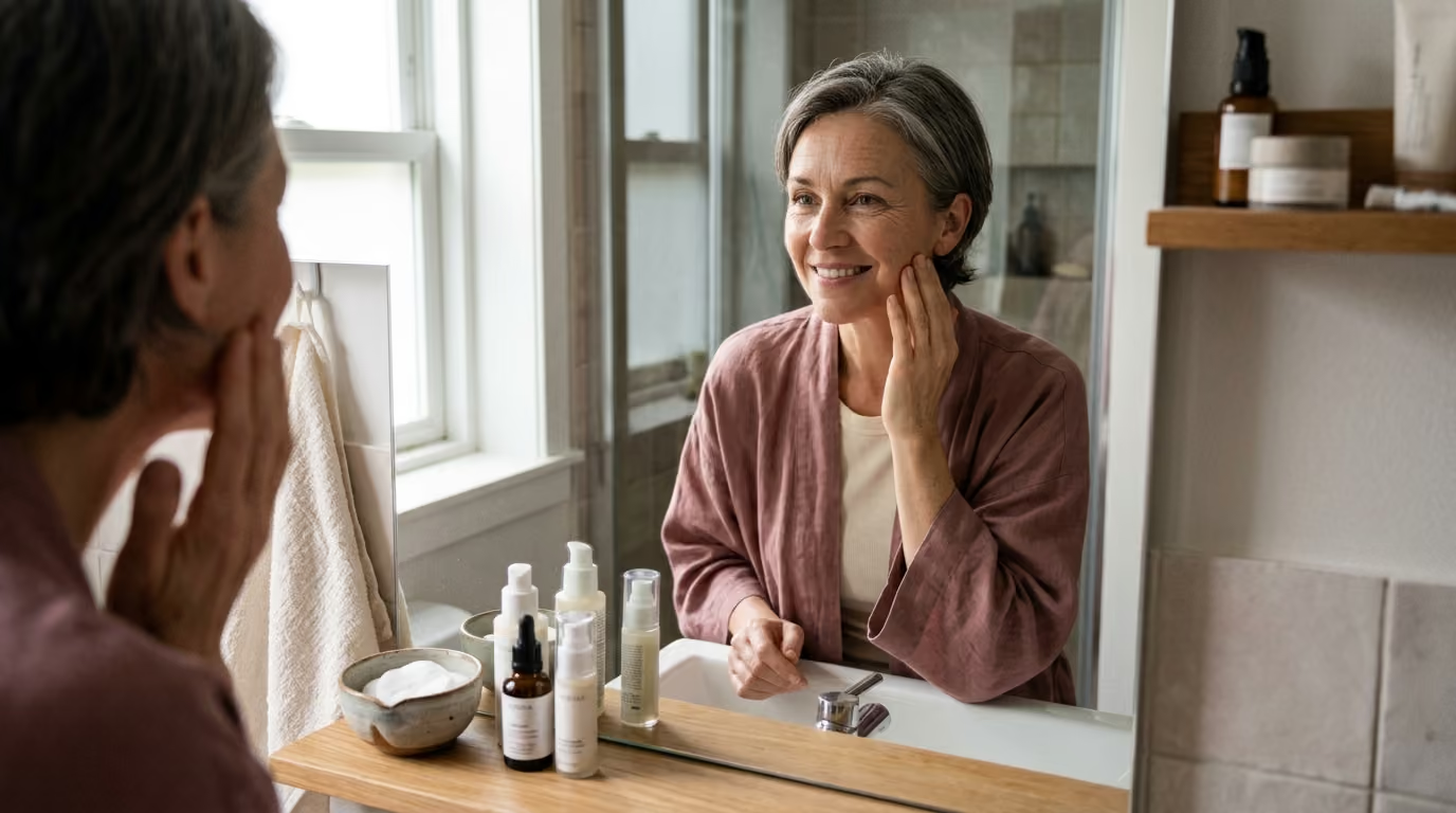

Advanced glycation end products modify dermal collagen and elastin structures, driving matrix stiffening, chronic inflammation.





Glycation is a natural chemical process where reducing sugars bond to functional proteins, lipids, and nucleic acids without enzymatic control. It is not an overnight catastrophe caused by eating a single sweet dessert, nor is it an avoidable biological event. Instead, glycation represents a slow, cumulative metabolic interaction that gradually modifies the physical structure and cellular behavior of long-lived tissues throughout the body.
Understanding how glycation influences skin longevity requires moving past simplified soundbites. The connection between sugar and skin aging involves cumulative glucose exposure, reactive carbonyl compounds, oxidative stress, ultraviolet radiation, and normal matrix turnover. By examining how advanced glycation end products form, attach to dermal proteins, and interact with cellular receptors, we can build a realistic, evidence-based approach to metabolic skin health.
To understand how glycation affects skin integrity, we must first separate it from essential biological mechanisms. The human body depends on carbohydrates for daily cellular energy and structural signaling. However, the way sugars interact with proteins depends entirely on whether enzymes guide the process.
Glycosylation is an enzyme-mediated, highly controlled biological process. During glycosylation, cells attach specific carbohydrate structures to proteins and lipids at precise molecular sites. This regulated modification is necessary for cell membrane stability, immune system recognition, and proper protein folding. Without glycosylation, human tissues could not function or repair themselves.
Glycation, by contrast, is completely nonenzymatic. It is a spontaneous chemical reaction that occurs when reducing sugars, such as glucose or fructose, encounter free amino groups on proteins, lipids, or DNA. Because no enzyme directs this reaction, it proceeds based on ambient sugar concentrations, temperature, time, and oxidative conditions. Over time, these unstable initial bonds undergo complex chemical rearrangements, yielding permanent structural modifications known as advanced glycation end products, or AGEs.
The initial stage of this transformation is part of the Maillard reaction, the same chemical cascade responsible for the browning of food during cooking. In human tissue, a reducing sugar reacts with a primary amine to form an unstable Schiff base. Within days, this base rearranges into a more stable intermediate known as an Amadori product.
Amadori products can undergo further oxidation, dehydration, and chemical fragmentation over weeks, months, or years. This progression generates highly reactive dicarbonyl intermediates, including methylglyoxal and glyoxal. These dicarbonyl compounds are substantially more chemically reactive than glucose itself. They rapidly attack nearby amino acid residues, culminating in the irreversible formation of diverse AGE structures.
Researchers have identified numerous distinct AGE structures in human tissues. Some of the most widely investigated variants include:
AGEs originate from both endogenous and exogenous sources. Endogenous AGEs form naturally inside the body as a byproduct of everyday glucose metabolism and cellular respiration. Their production rises during periods of sustained hyperglycemia or elevated systemic oxidative stress.
Exogenous AGEs, on the other hand, enter the body through the diet. They form in foods exposed to high-temperature, dry-heat cooking methods, such as roasting, frying, and grilling. While dietary AGEs are absorbed in the gastrointestinal tract, they are metabolized and cleared differently than endogenous compounds. Distinguishing between internally generated AGEs and dietary intake is essential when evaluating the actual drivers of long-term skin health.
The extracellular matrix of the dermis provides the structural foundation for visible skin firmness, tensile strength, and suppleness. Within this dense network, collagen accounts for the vast majority of dry tissue weight. Collagen fibers must withstand decades of mechanical tension, facial movement, and environmental exposure while maintaining a resilient, organized scaffold.
The primary reason dermal collagen suffers disproportionate glycation damage lies in its remarkable biological longevity. Unlike short-lived cellular proteins that turn over within hours or days, dermal collagen has an estimated biological half-life measured in years. Because these structural fibers remain embedded in the skin for extended periods, they experience prolonged, cumulative exposure to circulating glucose and reactive carbonyl intermediates.
When reducing sugars and reactive dicarbonyls react with the lysine and arginine residues of collagen, they do not merely sit on the surface of the fiber. Instead, they establish permanent intermolecular cross-links between adjacent collagen molecules. In healthy young skin, collagen fibers are organized in flexible, sliding bundles that glide smoothly over one another during movement. Glycation-induced cross-links act like rigid molecular bridges, locking these fibers together and preventing normal structural flexibility.
This structural immobilization alters the biomechanical profile of the skin through a distinct sequence:
Beyond changing physical flexibility, glycation disrupts the natural remodeling cycle of the dermis. Healthy tissue renewal depends on matrix metalloproteinases, or MMPs. These specialized enzymes identify, cleave, and clear worn extracellular proteins so that fibroblasts can synthesize fresh, well-organized replacements.
Glycated collagen demonstrates marked resistance to normal enzymatic degradation by MMPs. Because the chemical cross-links distort the natural cleavage sites, MMPs cannot efficiently break down damaged, glycated fibers. Consequently, the skin accumulates stiff, fragmented, and dysfunctional collagen that cannot be easily cleared.
This situation creates a biological paradox within the dermis. The extracellular matrix becomes denser and mechanically stiffer, yet it simultaneously loses structural integrity, cellular support, and self-renewal capacity. Fibroblasts residing within this rigid matrix receive distorted mechanical signals, leading to reduced synthesis of new procollagen. Over time, the balance between tissue breakdown, clearance, and regeneration shifts toward progressive structural disorganization.
While collagen provides tensile strength to resist pulling forces, dermal elastin provides the complementary mechanical quality of elastic recoil. Elastin fibers allow the skin to stretch under movement and snap back to its original resting state. In the absence of functional elastin, tissue stretches out, sags, and displays persistent surface slackness.
Just like collagen, dermal elastin is an exceptionally long-lived protein with minimal turnover in adult skin. Research shows that AGEs readily form cross-links within elastin fibers, directly modifying their unique coil-like molecular structure. When advanced glycation end products accumulate along elastin polymers, the fibers become brittle, calcified, and prone to mechanical fracturing.
To understand how these molecular changes alter visible skin quality, it helps to distinguish between distinct biomechanical terms:
Glycation creates a unique mechanical state where skin stiffness increases while functional elasticity decreases. An AGE-modified dermal matrix is harder and more resistant to initial stretching, yet it lacks the spring-like recoil needed to snap back cleanly. This combination accelerates visible tissue laxity, as the rigid yet unyielding structural network gradually yields to gravity and repetitive muscular pull.
Histological investigations of glycated skin reveal profound architectural disruption throughout the mid and deep dermis. Collagen bundles appear shortened, thinned, and randomly oriented rather than arranged in organized parallel patterns. The delicate microfibrillar network that anchors the epidermis to the underlying dermis also experiences glycation-related weakening.
These structural defects extend directly to tissue repair mechanisms. Glycated extracellular matrix components impair normal keratinocyte and fibroblast migration, leading to delayed wound closure, altered scar formation, and compromised barrier recovery. When the skin experiences a micro-injury, the surrounding rigid matrix fails to transmit the coordinated chemical and mechanical signals required for prompt healing.
Furthermore, dermal ground substance suffers secondary disruptions. The glycation of structural proteins alters their electrostatic charge and reduces their ability to bind water and hydrophilic proteoglycans. As a result, the dermis exhibits reduced internal hydration, leading to a duller surface reflection and a loss of visible firmness that topical moisturizers cannot easily resolve.
The physiological damage caused by glycation extends far beyond passive structural cross-linking. Advanced glycation end products function as active biological signaling molecules that alter cellular behavior throughout the skin. They achieve this primarily by binding to a specialized cell-surface protein called the receptor for advanced glycation end products, or RAGE.
RAGE belongs to the immunoglobulin superfamily of cell-surface molecules and is expressed on multiple cutaneous cell types, including dermal fibroblasts, epidermal keratinocytes, vascular endothelial cells, and resident immune cells. Under baseline conditions, RAGE expression remains relatively low. However, when circulating or matrix-bound AGEs bind to RAGE, they trigger an intracellular signal that upregulates RAGE expression, making the cell increasingly sensitive to further stimulation.
The biological consequences of AGE accumulation can be divided into two distinct yet overlapping pathways:
When AGEs engage RAGE on dermal fibroblasts, the cells shift away from balanced matrix synthesis toward a pro-oxidant, pro-inflammatory state. The sustained generation of reactive oxygen species depletes intracellular antioxidant reserves, such as glutathione, leaving the cell vulnerable to oxidative damage. This oxidative microenvironment directly damages mitochondrial DNA, impairing cellular energy production and accelerating cellular senescence.
This sequence establishes an ongoing biological cycle within the skin. Metabolic and environmental stressors generate reactive carbonyls, which convert into AGEs and bind to RAGE. The resulting oxidative stress and inflammatory signaling cause neighboring cells to release catabolic enzymes and inflammatory mediators. These inflammatory factors further damage extracellular proteins and disrupt cellular repair, creating additional substrate for future glycation reactions.
Understanding this dual mechanism clarifies why glycation is a central topic in skin longevity and healthy aging research. Managing the impact of glycation requires addressing both the physical integrity of structural fibers and the cellular inflammatory pathways that drive chronic tissue stress.
A frequent misconception in aesthetic discussions is that glycation and photoaging represent two entirely separate pathways of skin aging. In reality, ultraviolet radiation and glycation operate as interconnected catalysts that compound one another's destructive effects on the dermal matrix.
Ultraviolet A (UVA) radiation penetrates deep into the dermis, where it generates high levels of reactive oxygen species through interactions with endogenous photosensitizers. Ultraviolet B (UVB) radiation primarily affects the epidermis, triggering direct DNA mutations and acute inflammatory cascades. Both forms of radiation amplify glycation biology through distinct chemical interactions.
Research demonstrates that solar radiation accelerates the formation of advanced glycation end products in sun-exposed tissue. UVA exposure generates substantial oxidative stress, which drives the lipid peroxidation and glycoxidation reactions required to convert intermediate Amadori products into permanent AGEs like carboxymethyl-lysine. Consequently, biopsies from photoexposed facial skin consistently show higher concentrations of CML and pentosidine than sun-protected skin from the same individual.
Furthermore, glycated proteins within the extracellular matrix act as endogenous photosensitizers. When UVA photons strike an AGE-modified collagen fiber, the modified molecule absorbs the energy and enters an excited electronic state. As it returns to baseline, it transfers energy to ambient molecular oxygen, generating singlet oxygen and superoxide radicals.
This photodynamic reaction causes localized collateral damage:
The interaction between sunlight and glycation also contributes to the visible discoloration associated with mature skin. Advanced glycation end products possess characteristic yellow-brown chromophores that absorb light in the blue spectrum. When these pigmented cross-links accumulate within photoexposed elastotic tissue, they impart a dull, yellowish or sallow undertone to the complexion.
This deep biological interaction reinforces a foundational principle of evidence-based skin health: nutritional and metabolic adjustments cannot compensate for unshielded sun exposure. Photoprotection remains an indispensable foundation for mitigating the oxidative environment that allows advanced glycation end products to thrive.
Public discussions about diet and skin aging frequently rely on exaggerated claims, suggesting that eating a single sugary snack instantly degrades facial collagen. Scientific evaluation requires examining human clinical data and epidemiological studies with greater precision and nuance.
Large-scale observational research provides important context regarding dietary intake and tissue AGE accumulation. The Rotterdam Study, a major population-based investigation analyzing 2,515 participants, evaluated the relationship between estimated dietary AGE intake and measured skin autofluorescence. The researchers measured dietary intake of specific markers, including carboxymethyl-lysine (CML), carboxyethyl-lysine (CEL), and methylglyoxal-derived hydroimidazolone (MGH1).
The findings from the Rotterdam Study revealed complex, nuanced outcomes:
These findings highlight why we must separate dietary sugar intake from tissue glycation burden. When a person consumes carbohydrates, the digestive tract breaks them down into simple monosaccharides, which are absorbed into the bloodstream and processed by the liver, skeletal muscle, and adipose tissue. The extent to which dietary carbohydrates contribute to tissue glycation depends on systemic glycemic regulation, insulin sensitivity, physical activity, and baseline metabolic health.
Authoritative global health bodies provide clear parameters for sugar consumption. The World Health Organization strongly recommends that adults and children reduce their daily intake of free sugars to less than 10% of total energy intake. The WHO further notes that a reduction to below 5% of total energy intake may provide additional health benefits.
It is critical to interpret these public health guidelines correctly:
Whole fruits contain dietary fiber, water, micronutrients, and bioactive polyphenols that slow gastric emptying and blunt postprandial glucose excursions. Furthermore, many plant polyphenols possess natural antioxidant properties that help neutralize reactive carbonyl species in preclinical models. Conflating a fresh apple or bowl of berries with a glass of refined corn syrup ignores the metabolic context of digestion and systemic nutrient delivery.
Human skin aging reflects total glycemic exposure over years rather than transient post-meal fluctuations. A dietary pattern that supports metabolic stability, emphasizes minimally processed whole foods, and provides adequate micronutrients creates an internal environment that naturally moderates glycation kinetics without requiring extreme restriction.
Because glycation is driven by ambient glucose concentrations and oxidative stress, systemic metabolic conditions dramatically alter the rate at which advanced glycation end products form in dermal tissue. Examining clinical disease models provides clear insight into the physiological drivers of tissue glycation.
Persistent hyperglycemia, the hallmark of poorly controlled type 1 and type 2 diabetes, creates an environment of elevated glycation pressure. When circulating blood glucose remains elevated over long periods, the rate of nonenzymatic Schiff base formation rises proportionally. Consequently, individuals with long-standing diabetes demonstrate substantially higher concentrations of advanced glycation end products in skin biopsies and noninvasive optical measurements.
Clinical studies tracking skin autofluorescence in diabetic populations have established several key observations:
Chronic kidney disease represents another critical clinical context that influences glycation dynamics. The kidneys play an essential role in filtering and clearing circulating advanced glycation end products and reactive dicarbonyl intermediates from the bloodstream. When renal function declines, the physiological clearance of these compounds is impaired, leading to elevated systemic retention and accelerated tissue deposition.
This renal connection explains why individuals with chronic kidney disease frequently present with elevated skin autofluorescence regardless of their dietary sugar intake. If the body cannot efficiently excrete metabolic byproducts, AGE precursors accumulate in long-lived tissues. This reality underscores why skin autofluorescence scores cannot be interpreted solely as a reflection of personal lifestyle or cosmetic habits.
Beyond diagnosed disease states, subclinical metabolic dysfunction, such as insulin resistance and chronic low-grade inflammation, influences glycation biology. When skeletal muscle and liver cells become resistant to insulin, postprandial glucose spikes become taller and remain elevated longer. This prolonged exposure increases the likelihood of reactive carbonyl formation and cellular oxidative stress.
Managing metabolic health through regular physical exercise, balanced macronutrient distribution, restorative sleep, and routine medical oversight represents the most scientifically sound approach to moderating glycation. Protecting the structural matrix of the skin requires maintaining the health of the metabolic and vascular systems that supply it.
As scientific interest in longevity and tissue health has grown, researchers have sought reliable methods to quantify advanced glycation end products in living human tissue. Measuring dermal glycation offers valuable insight into biological aging and systemic metabolic stress, but the technologies used have important capabilities and limitations.
The gold standard for quantifying tissue AGEs is biochemical analysis of full-thickness skin biopsies using techniques like liquid chromatography-tandem mass spectrometry (LC-MS/MS) or enzyme-linked immunosorbent assays (ELISA). While highly accurate and capable of quantifying specific nonfluorescent structures such as CML and CEL, tissue biopsies are invasive, cause localized scarring, and are impractical for routine screening.
To address these challenges, researchers developed noninvasive optical measurement systems, most notably skin autofluorescence (SAF) using devices like the AGE Reader. This technology relies on the optical properties of specific advanced glycation end products that fluoresce when excited by light of specific wavelengths.
The AGE Reader illuminates a small patch of skin, typically on the inner forearm, with ultraviolet A light ranging from 300 to 420 nanometers. Fluorescent AGEs embedded within the dermal collagen, such as pentosidine and crossline, absorb this optical energy and emit fluorescent light at wavelengths between 420 and 600 nanometers. Photodetectors measure the intensity of the emitted light relative to the reflected excitation light, calculating an arbitrary unit score that reflects the concentration of fluorescent dermal AGEs.
Clinical validation studies demonstrate that skin autofluorescence correlates with collagen-linked fluorescence and specific AGE concentrations extracted from human skin biopsies. SAF has proven valuable in clinical research for stratifying cardiovascular risk and assessing long-term cumulative metabolic exposure in populations with diabetes and renal impairment.
However, skin autofluorescence has meaningful technical and clinical limitations:
Understanding these technical parameters allows us to appreciate noninvasive optical tools for what they are: helpful research instruments that measure specific fluorescent markers, rather than infallible diagnostic tools for individual skin appearance.
Addressing the biological impact of glycation requires a practical, multifaceted framework. Rather than searching for quick fixes or adopting extreme dietary rules, the most effective approach combines sensible nutrition, photoprotection, evidence-backed topical ingredients, and overall metabolic care.
The most direct way to moderate endogenous AGE formation is to maintain healthy blood glucose regulation and insulin sensitivity over time.
Because ultraviolet radiation acts as a powerful catalyst for oxidative stress and AGE cross-linking, photoprotection remains essential for supporting the dermal matrix.
Dietary AGEs form most readily when foods, particularly proteins and fats, are exposed to high-temperature, dry-heat cooking methods. Shifting cooking habits can help lower exogenous AGE intake without sacrificing meal quality.
A variety of cosmetic and dermatological ingredients have been evaluated for their potential to support glycated or mature skin.
In pharmacological research, compounds known as "AGE breakers" (such as early experimental agents like ALT-711 or alagebrium) were designed to chemically cleave established cross-links in collagen and vascular tissue. While these agents demonstrated efficacy in preclinical animal models, human clinical trials yielded mixed results, and several compounds were discontinued due to safety concerns or lack of clear clinical benefit.
Similarly, aminoguanidine showed strong capacity to trap reactive carbonyls in laboratory models, but its clinical development was halted due to adverse side effects in diabetic trials. Currently, there is no validated topical cosmetic ingredient or dietary supplement proven to erase established collagen cross-links in living human skin. Preventing future modification and supporting natural matrix turnover remain the most scientifically grounded approaches.
To learn more about optimizing your skincare and lifestyle framework, explore our comprehensive guides on lifestyle, recovery, and environmental aging alongside nutrition and beauty from within.
The intersection of nutrition and dermatology frequently generates sensationalized headlines. Separating biological fact from marketing fiction is essential for maintaining a healthy relationship with food and skincare.
Reality: Glycation is a slow, concentration-dependent chemical process that unfolds over weeks, months, and years. Transient, normal elevations in blood glucose following a meal do not instantly lock dermal collagen fibers into rigid cross-links. Cumulative, multi-year glycemic control and baseline oxidative stress dictate long-term AGE accumulation.
Reality: Whole fresh fruits are rich in dietary fiber, vitamins, water, and protective polyphenols. The fiber in whole fruit slows carbohydrate absorption, preventing sharp glucose spikes. Scientific guidance from the World Health Organization targets added free sugars and liquid sweeteners, not whole, intact fruits.
Reality: Endogenous glycation occurs continuously as part of normal human metabolism. Furthermore, environmental factors such as ultraviolet radiation, chronic psychological stress, cigarette smoking, and air pollution accelerate oxidative stress, driving reactive carbonyl formation independently of diet.
Reality: Once stable, covalent advanced glycation end products cross-link deep within dermal collagen, they are exceptionally difficult to cleave without damaging the underlying protein scaffold. Skincare ingredients like carnosine and antioxidants can help neutralize reactive intermediates and protect newly synthesized collagen, but they do not dissolve established structural cross-links.
Reality: Extreme dietary restriction does not stop aging. Severe carbohydrate deprivation can increase systemic cortisol, impair thyroid function, and lead to micronutrient imbalances that compromise skin barrier repair and dermal hydration. Balanced, sustainable nutrition supports long-term health far more effectively than nutritional extremes.
Regular physical activity improves whole-body insulin sensitivity and increases glucose uptake by skeletal muscle through non-insulin-dependent pathways. By helping maintain stable baseline blood glucose concentrations and lowering systemic oxidative stress, consistent exercise indirectly moderates the internal conditions that drive endogenous AGE formation.
Cooking methods substantially influence the exogenous AGE content of food. Preparing lean proteins and vegetables with moist-heat methods, such as steaming, poaching, or stewing, generates significantly fewer dietary AGEs than high-temperature frying, broiling, or charring. However, overall dietary quality, nutrient variety, and metabolic health remain the primary drivers of tissue longevity.
There is no conclusive biological evidence indicating that melanin levels increase the physiological rate of collagen glycation. However, optical measurement devices like the AGE Reader face technical limitations in individuals with Fitzpatrick skin types V and VI because high epidermal melanin absorbs the light signals, which can cause measurement errors.
Ingested collagen supplements are broken down during digestion into individual amino acids and small bioactive dipeptides, such as prolyl-hydroxyproline. These absorbed peptides enter the bloodstream and can stimulate dermal fibroblasts to produce fresh collagen and hyaluronic acid. However, oral collagen peptides cannot seek out, chemically cleave, or reverse existing AGE cross-links in established dermal fibers.
Stay connected for research and practical guidance on skin, hair, collagen, nutrition and beauty longevity. Clear ideas for people who want to understand how appearance changes with age and make better-informed choices over time.


Understand your skin, hair and body better without chasing every new trend, treatment or promise.
explore the Blog