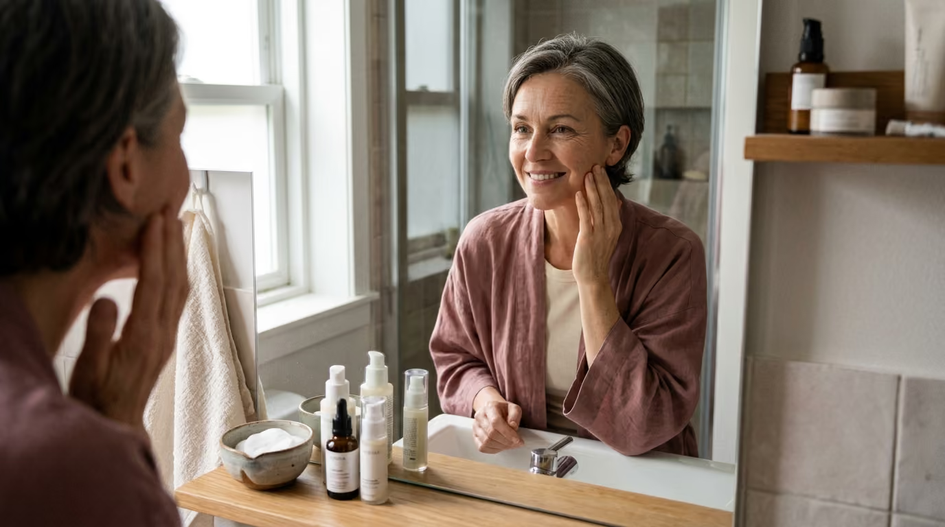

When standard skincare advice fails your tone, genetic differences in melanin, collagen density, and hormones explain how personalized longevity routines.





Skin longevity is not a uniform biological timeline. It is not a fixed countdown determined solely by the year on your birth certificate. Instead, visible skin aging reflects the continuous interaction of your genetic background, baseline pigmentation, deep anatomical architecture, hormonal milestones, and environmental exposures.
Popular beauty culture often reduces this complex biological reality to simple generalities. Lighter skin is frequently warned about premature fine lines. Darker skin is often told that it possesses built-in immunity to sun damage. Both narratives oversimplify human biology and lead to ineffective or potentially harmful routines.
A personalized understanding of skin longevity requires looking past broad labels. In this resource, we examine the molecular genetics of pigmentation, structural differences across diverse skin phenotypes, the impact of hormonal shifts, and the nuanced risks of clinical treatments. By understanding how these variables intersect, you can build a targeted, long-term approach to skin health that honors your unique biological profile.
Visible skin aging is governed by both internal biological clocks and external lifestyle factors. The interaction between these forces varies significantly across different genetic backgrounds and skin tones.
Understanding these biological findings helps clarify why two individuals of the exact same age often require entirely different approaches to maintain healthy skin over time.
Skin pigmentation is one of the most genetically variable traits in human biology. It evolved primarily as an evolutionary adaptation to regional ultraviolet radiation levels across the globe. Rather than relying on a single genetic switch, melanin production is governed by a distributed polygenic network.
The melanocortin 1 receptor gene, known as MC1R, plays a central role in controlling melanocyte signaling. It determines the balance between dark brown eumelanin and yellow-red pheomelanin. Other vital genes include TYR, which encodes the essential enzyme tyrosinase, alongside TYRP1 and DCT. Specific genetic variants, such as the TYR variant rs1126809 and the IRF4 variant rs12203592, directly modulate melanin synthesis, tanning efficiency, freckling patterns, and cellular sensitivity to environmental light.
Scientists separate skin color into two distinct categories: constitutive pigmentation and facultative pigmentation. Constitutive pigmentation refers to your baseline, genetically programmed skin tone without environmental exposure. Facultative pigmentation describes the deepened pigment acquired after exposure to ultraviolet radiation, systemic hormones, inflammation, or physical trauma.
Eumelanin provides natural photoprotection by absorbing and dissipating ultraviolet energy before it can create extensive DNA mutations. This natural defense explains why the classic signs of light-induced aging, such as deep wrinkling, often appear later in richly pigmented complexions. However, this protection is incomplete. Ultraviolet rays still induce oxidative stress, cellular senescence, and DNA damage in melanated skin cells.
Furthermore, melanocytes in darker skin are biologically reactive. When exposed to inflammatory triggers or physical injury, they frequently overproduce pigment. This reaction leads to persistent dyschromia and post-inflammatory hyperpigmentation. To learn more about cellular health and longevity, read our detailed guide to skin longevity and healthy aging.
Skin longevity is deeply tied to the structural integrity of the dermis and underlying tissues. Dermal fibroblasts synthesize collagen and elastin, which form the supportive extracellular matrix of the skin. Research shows measurable differences in this structural architecture across different ancestral backgrounds.
Histological analyses show that individuals of Asian and African descent frequently possess a thicker, more compact dermis than individuals of European descent. In these compact dermal layers, collagen bundles are densely woven, and fibroblast activity remains robust across middle age. Additionally, advanced imaging reveals that the dermal-epidermal junction exhibits greater structural height and cellular adhesion in African American cohorts compared to Caucasian cohorts.
These structural differences explain why the visual patterns of aging vary widely across populations. In lighter skin types with thinner dermal layers, loss of elasticity and superficial rhytides frequently emerge as the primary early concern. In more richly pigmented skin, the compact dermal matrix provides natural resistance against superficial crepiness.
However, structural firmness does not prevent facial aging altogether. Facial aging is a three-dimensional process that spans every anatomical plane:
Defining aging purely by wrinkle depth ignores these deeper changes. A person may have few visible wrinkles yet show substantial midface volume descent or pronounced textural changes. To understand how structural protein matrices degrade over time, explore our analysis of collagen and structural aging.
Visible skin changes represent the cumulative impact of internal biological aging, external exposures, and hormonal transitions. The scientific community describes this collection of lifetime environmental factors as the skin exposome.
Ultraviolet radiation remains the most prominent driver of external skin damage. Solar-simulated radiation and UVB wavelengths strongly trigger melanogenesis-related gene expression. Notably, clinical data demonstrates that UV-induced pigment gene expression is significantly stronger in older skin than in younger skin. This heightened reactivity explains why older individuals often develop stubborn, uneven dark spots following routine sun exposure.
Visible light, which spans wavelengths between 400 and 700 nanometers, also plays a critical role in pigmentary disorders. While visible light causes minimal pigment response in fair skin, it triggers intense, long-lasting hyperpigmentation in darker complexions. Research indicates that visible light induces reactive oxygen species and stimulates stable tyrosinase complexes in melanocytes. Standard mineral or chemical sunscreens that only block UVA and UVB rays fail to protect against these visible wavelengths.
Urban air pollution acts as an additional environmental accelerator. Experimental research investigating combined exposure to ultraviolet light and urban particulate matter showed marked epidermal thinning and barrier disruption. These particulate pollutants triggered melanin leakage into the underlying dermis. This biological response closely mirrors the tissue changes observed in clinical post-inflammatory hyperpigmentation and solar lentigines.
Simultaneously, internal hormonal shifts dramatically alter tissue longevity. Estrogen directly stimulates dermal fibroblasts to produce collagen, elastin, and hyaluronic acid. During the menopausal transition, the rapid decline in circulating estrogen causes a pronounced loss of structural tissue.
Clinical investigations show that skin collagen decreases by approximately 30 percent during the initial five years following menopause. Over the subsequent 15 years, collagen levels decline by an additional 2.1 percent each year. This loss correlates directly with postmenopausal duration rather than chronological age. A 52-year-old individual undergoing rapid hormonal transitions may experience faster tissue thinning and dryness than a 58-year-old with stable hormone levels.
In aesthetic medicine and clinical dermatology, a standard treatment protocol can produce vastly different outcomes across different skin tones. The primary reason for this variation is the biological link between inflammation and melanocyte activation.
In skin of color, treatments that cause epidermal disruption, deep thermal heat, or physical trauma can provoke severe post-inflammatory hyperpigmentation. This creates a challenging clinical situation where an aggressive procedure designed to correct tone or texture instead generates new, long-lasting pigmentation.
Energy-based devices such as lasers illustrate these treatment risks. Ablative resurfacing lasers and deep chemical peels carry substantial safety concerns for individuals with Fitzpatrick phototypes IV through VI. Clinical reviews indicate that repeated procedural laser applications produce post-inflammatory hyperpigmentation in roughly 11 to 17 percent of patients with skin of color. In chart reviews of traditional carbon dioxide laser resurfacing, hyperpigmentation occurred in 37 percent of patients across all skin tones, with the highest rates found in darker complexions.
Because of these procedural risks, topical interventions remain the first line of care for managing dyschromia. A 12-week randomized clinical trial in individuals with Fitzpatrick types III through VI compared a standard four percent hydroquinone system with a non-hydroquinone brightening regimen containing targeted botanical brighteners and retinoids. Both systems achieved statistically significant improvements in overall hyperpigmentation, with the hydroquinone group showing slightly faster clearance.
Both treatment groups maintained low irritation rates, which was critical to their overall success. If a topical active ingredient causes persistent redness or peeling, the resulting inflammation can stimulate melanocytes and worsen the underlying discoloration. For readers exploring cellular signaling and modern skincare formulation, our overview of beauty science offers deeper insights into topical delivery systems.
When we first started reviewing clinical trials on structural skin aging, I was struck by how often popular media misinterpreted the data. A study showing a minor increase in skin elasticity was suddenly headlined as a total reversal of aging. It made me realize how desperately consumers need a translator for beauty science, someone who can say exactly what a study proves and what it does not prove.
Current genetic and dermatological research has clear limitations that consumers should understand:
Most direct-to-consumer genetic panels analyze a small selection of single nucleotide polymorphisms. While carrying a specific variant in the MC1R or IRF4 gene influences your sensitivity to ultraviolet light, it cannot account for your daily sun protection habits, sleep quality, local air quality, or barrier health. Genetic data provides broad risk probabilities rather than fixed clinical outcomes.
Furthermore, dermatological literature has historically relied on broad ancestral labels like Caucasian, Asian, and Black. These expansive categories contain immense internal genetic, geographic, and phenotypic variation. Treating any population as a uniform group ignores the diverse range of skin tones, dermal structures, and inflammatory responses found within it.
Creating an effective skincare strategy requires looking at your individual phenotype, environmental exposures, and tissue health. Rather than following broad trends, you can adopt a systematic approach to support long-term skin health.
Document your baseline skin characteristics instead of relying solely on ethnic background. Evaluate your Fitzpatrick phototype, baseline skin tone, and your personal history of sunburns versus tanning. Record whether you develop dark marks after acne breakouts, bug bites, or minor scratches, and note any personal tendency toward hypertrophic scarring or keloids.
Determine which biological process requires the most attention:
Targeting your primary biological concern prevents product overload and reduces the risk of ingredient-induced irritation.
Examine your daily environment and lifestyle habits. Account for regional ultraviolet intensity, daily commuting time, outdoor hobbies, and occupational exposure to heat or urban air pollution. Review photosensitizing medications and assess how consistently you apply sun protection throughout the day. To learn how daily habits influence skin vitality, read our guide on lifestyle, recovery, and environmental aging.
Before undergoing clinical treatments such as chemical peels, microneedling, or energy-based lasers, evaluate your risk of hyperpigmentation. High-risk indicators include having Fitzpatrick phototypes III through VI, active melasma, a history of post-inflammatory marks, or recent sun exposure. High-risk skin benefits from pre-treatment skin priming with topical tyrosinase inhibitors, conservative energy settings, and small test patches.
Maintaining barrier function is essential across all skin tones, especially during periods of hormonal transition or corrective pigment care. A compromised stratum corneum allows irritants to penetrate the skin, accelerating low-grade inflammation and triggering excess pigment production. Support barrier lipids by using gentle, non-foaming cleansers and moisturizers formulated with ceramides, physiological fatty acids, and cholesterol.
Evaluate your skin's health using comprehensive criteria rather than focusing solely on fine lines. Track overall skin tone uniformity, tissue hydration, barrier recovery speed, tactile smoothness, and personal comfort. Improving skin longevity means cultivating resilient, well-functioning tissue over the course of your life. For a broader overview of healthy aging, explore our general skin care research.
Widespread skincare assumptions often lead people to choose inappropriate products or overlook essential preventive habits. Reviewing these common beliefs through a scientific lens helps clarify what the evidence actually supports.
The belief that darker skin does not require sun protection remains one of the most common and misleading assumptions in beauty culture. While higher eumelanin levels provide meaningful defense against sunburn, ultraviolet and visible light still generate free radicals and drive uneven pigmentation across all phototypes.
Another widespread misconception is that faster cellular turnover requires aggressive resurfacing. In skin prone to hyperpigmentation, aggressive exfoliation frequently backfires by causing inflammatory trauma that triggers melanocytes to produce more pigment. A calm, measured approach focused on barrier preservation consistently delivers safer, more predictable results.
Finally, assuming that skin discoloration is purely a cosmetic concern can delay necessary medical care. Pigmented lesions, sudden patches of melasma, or dark marks on the palms, soles, and nail beds require careful clinical evaluation. Serious conditions like acral lentiginous melanoma can develop across all skin tones and require prompt medical attention.
Acral lentiginous melanoma is a distinct form of melanoma that develops on non-sun-exposed areas, including the palms of the hands, soles of the feet, and beneath the nail beds. Unlike cutaneous melanomas linked to chronic ultraviolet radiation, acral melanoma arises independently of sun exposure. It represents a larger proportion of total melanoma diagnoses in Black, Hispanic, and Asian populations. Because of the misconception that darker skin does not develop skin cancer, these lesions are often identified at later, more advanced stages. Regular self-examinations should always include checking the feet, nail beds, and palms for new, changing, or irregular dark spots.
Standard chemical and mineral sunscreens are designed to filter ultraviolet radiation, spanning wavelengths from 290 to 400 nanometers. However, visible light, which ranges from 400 to 700 nanometers, also penetrates the skin and stimulates melanocytes in individuals with Fitzpatrick phototypes III through VI. Non-tinted mineral and chemical filters do not adequately block these visible wavelengths. Tinted sunscreens contain iron oxides and pigmentary titanium dioxide, which create a physical barrier that reflects visible light. Clinical research confirms that using tinted formulations containing iron oxides helps prevent melasma flare-ups and post-inflammatory dark marks far more effectively than untinted sunscreens.
Clinical treatments for individuals with reactive or deeply pigmented skin should follow a conservative, step-by-step protocol. Dermatologists frequently recommend preparing the skin with topical tyrosinase inhibitors, such as azelaic acid, tranexamic acid, or low-dose hydroquinone, for two to four weeks prior to an in-office procedure. In-clinic providers avoid aggressive, ablative laser settings in favor of conservative, lower-fluence options, longer pulse durations, and superficial chemical peels. Performing a discreet test spot behind the ear or along the jawline helps clinicians assess personal tissue reactivity before treating the entire face.
A thicker, more compact dermis provides structural resistance against superficial skin crepiness and early fine lines, but it does not stop facial aging. Age-related changes occur throughout every anatomical layer of the face. Over time, deep and superficial fat compartments shift, facial retaining ligaments lose mechanical tone, and facial bones undergo gradual structural resorption. These deeper shifts lead to midface volume loss, deepening of the nasolabial folds, and lower-face skin laxity, even when the skin's surface remains relatively smooth and free of wrinkles.
Stay connected for research and practical guidance on skin, hair, collagen, nutrition and beauty longevity. Clear ideas for people who want to understand how appearance changes with age and make better-informed choices over time.


Understand your skin, hair and body better without chasing every new trend, treatment or promise.
explore the Blog