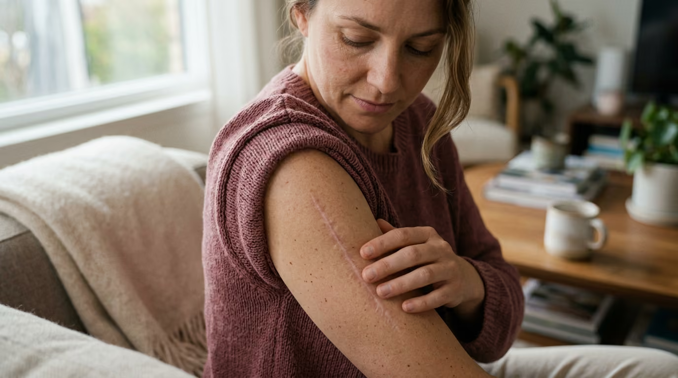

Seventy to eighty percent of dermal dry weight consists of collagen, governing wound healing phases, matrix remodeling, and varied scar formations.





Popular beauty marketing often suggests that scars are simply excess collagen deposits that need to be dissolved, or that stimulating new collagen will effortlessly repair any uneven texture. Both ideas misrepresent how human skin heals. A scar is not an arbitrary surplus of protein. It is an organized biological replacement produced when a wound disrupts the deeper dermal architecture.
Adult wound healing prioritizes rapid closure and tensile integrity over visual perfection. When an injury extends past the superficial epidermis into the dermis, the body builds a dense, temporary scaffold to restore mechanical strength. Understanding the biological timeline of this process clarifies why scars look, feel, and behave differently across various body sites and skin types.
Analyzing how the extracellular matrix responds to injury allows you to distinguish between normal maturation and pathological tissue changes. This guide provides an evidence-based examination of collagen deposition, enzymatic matrix remodeling, and the structural differences between normal, hypertrophic, keloid, and atrophic scars.
The skin functions as a multi-layered barrier against environmental stress, water loss, and physical trauma. The outer layer, known as the epidermis, provides the primary permeability barrier through tightly packed keratinocytes and lipid bilayers. Directly beneath lies the dermis, a thick connective tissue layer that gives the skin its structural integrity, elasticity, and tensile strength.
The dermal extracellular matrix is an intricate, water-rich environment composed of structural proteins, glycoproteins, proteoglycans, and hyaluronic acid. Specialized cells called fibroblasts inhabit this space, continuously producing and maintaining these structural elements. Collagen represents the single most abundant protein within this matrix, comprising roughly seventy to eighty percent of the dry weight of human skin.
Dermal collagen provides far more than superficial skin firmness. It creates a three-dimensional mechanical framework that couples physical forces between cells and their surroundings. This matrix supports blood vessels, nerve endings, and hair follicles while acting as a physical highway for immune cells during tissue repair. Without an intact collagen architecture, the skin loses its ability to resist deformation, shear stress, and tearing.
In healthy, uninjured adult skin, collagen fibers form an interlocking, three-dimensional basketweave pattern. This randomized orientation permits the skin to stretch and recover smoothly in multiple directions. The extracellular matrix also contains elastin fibers, which supply elastic recoil, and ground substance, which binds moisture to cushion mechanical shocks. When an injury penetrates deep into the dermis, this delicate basketweave architecture is destroyed, forcing fibroblasts to deploy an emergency repair protocol.
Wound healing follows an overlapping biological sequence: inflammation, proliferation, and remodeling. Each phase features specific cellular populations, biochemical signals, and extracellular matrix changes. A smooth transition through each step is essential for normal tissue repair.
The process begins immediately upon physical injury. Platelets aggregate at the damaged site, forming a fibrin clot that stops active bleeding and establishes a provisional physical plug. These activated platelets release chemotactic signals, including platelet-derived growth factor and transforming growth factor-beta, which draw inflammatory cells to the wound bed.
Neutrophils arrive within hours to destroy invading bacteria and clear cellular debris through phagocytosis. Within two to three days, circulating monocytes migrate into the tissue and differentiate into wound macrophages. Macrophages act as the directors of repair, shifting from an inflammatory clean-up role to a pro-repair state. They clear remaining debris, release cytokines, and instruct fibroblasts to begin building new tissue.
Inflammation is a necessary protective phase, but it must remain strictly time-limited. If infection, mechanical irritation, or foreign material causes inflammation to persist, the signaling cascade becomes dysregulated. Chronic, unresolved inflammation is one of the primary drivers of pathological scarring.
Beginning around the third day and lasting several weeks, the proliferative phase focuses on filling the physical gap and restoring tissue continuity. Endothelial cells sprout from existing capillaries to form a dense microvascular network in a process known as angiogenesis. This new, highly vascularized bed is called granulation tissue, which appears bright pink or red and bleeds easily upon contact.
Fibroblasts migrate into this granulation tissue and rapidly synthesize large amounts of type III collagen and glycosaminoglycans. Type III collagen contains smaller, more flexible fibers than mature dermal collagen, allowing for quick deposition and cellular movement across the wound. During this window, specialized fibroblasts differentiate into myofibroblasts under the influence of mechanical tension and growth factors.
Myofibroblasts express alpha-smooth muscle actin, granting them contractile capabilities similar to smooth muscle cells. They grip the surrounding matrix fibrils and pull the wound edges closer together, reducing the total surface area requiring cellular coverage. Keratinocytes simultaneously migrate across this moist bed to re-establish the epidermal barrier. Once the wound is fully closed, myofibroblasts typically undergo programmed cell death, known as apoptosis.
The remodeling phase begins roughly three weeks after the initial injury and continues for twelve months or longer. During this extended period, the body systematically breaks down the provisional matrix and replaces it with a more durable structural framework.
Matrix metalloproteinases, which are zinc-dependent enzymes secreted by fibroblasts and immune cells, degrade the temporary type III collagen. Concurrently, fibroblasts synthesize thicker, more mechanically robust type I collagen. As this replacement unfolds, the ratio of type I to type III collagen gradually normalizes toward the baseline seen in healthy adult skin.
The newly synthesized type I collagen fibers are organized into dense parallel bundles, rather than the original basketweave network. Covalent cross-links form between individual collagen molecules, steadily increasing the physical strength of the repair. Microvessels regress, causing the initial redness to fade, while the total cellularity of the tissue declines.
Every deep cut or surgical incision in an adult yields a scar, but the biological characteristics of that scar can vary substantially. Differences in inflammation, cellular apoptosis, and matrix turnover produce distinct clinical presentations.
A normal scar represents an efficient, self-limiting healing response. The repair stays confined to the original injury margin and undergoes an orderly progression from vascular and firm to pale and soft.
During early remodeling, a normal scar may appear raised, red, and mildly firm due to active blood vessels and provisional collagen. Over several months, as enzyme activity balances collagen production, the scar flattens and softens. While a normal scar will never fully replicate the original dermal architecture, it remains asymptomatic and functionally stable.
Hypertrophic scars are raised, rigid, and erythematous lesions that remain strictly within the boundaries of the original wound. They often develop within weeks of a burn, deep trauma, or high-tension surgical incision.
The underlying biology of hypertrophic scarring involves sustained, low-grade inflammation and elevated fibroblast activity. A primary feature is the failure of myofibroblasts to undergo normal apoptosis once the wound closes. Because these contractile cells persist, they continue to produce alpha-smooth muscle actin and deposit excess extracellular matrix.
Research indicates that hypertrophic scars synthesize roughly three times more collagen than normal healing skin. The collagen bundles in these lesions form dense, nodular patterns parallel to the skin surface. Patients frequently experience persistent itching, local pain, and a feeling of tissue tightness. Over several years, hypertrophic scars may undergo partial spontaneous regression, though they rarely disappear without medical management.
Keloids represent a distinct fibroproliferative disorder characterized by aggressive, continuous matrix accumulation. Unlike hypertrophic scars, keloids actively invade surrounding healthy tissue, expanding well beyond the original boundaries of the injury.
Keloid fibroblasts exhibit an abnormal, hyper-responsive phenotype that behaves similarly to a benign proliferative process. These cells produce high levels of growth factors, resist normal apoptotic signals, and demonstrate impaired matrix degradation. Some laboratory analyses estimate that keloid tissue synthesizes up to twenty times more collagen than uninjured dermal tissue.
Keloids rarely regress on their own and can continue growing for years following a minor skin injury. They are particularly common after earlobe piercings, sternal incisions, burns, and severe acne. While keloids can develop in any individual, clinical data shows a significantly higher incidence in darker Fitzpatrick skin types (types IV through VI). Genetic susceptibility, personal history, and anatomical tension all contribute to keloid development.
Atrophic scars present as sunken, depressed depressions below the plane of the surrounding skin. Rather than an overproduction of extracellular matrix, atrophic scars result from a net destruction or inadequate replacement of dermal collagen.
These scars are most frequently caused by localized, severe inflammation from inflammatory acne or varicella. When an inflamed sebaceous follicle ruptures deep within the dermis, an intense immune reaction destroys collagen, elastin, and subcutaneous adipose tissue. If the subsequent proliferative phase fails to synthesize sufficient new matrix to replace what was lost, the epidermis heals over an underlying structural void.
Dermatology classifies atrophic acne scars into three major morphological subtypes:
Rolling scars are frequently held down by dense fibrous tethering bands in the deep dermis, pulling the epidermis downward. Addressing rolling scars often requires physically severing these bands rather than simply applying topical therapies. Those seeking deeper insights into how the dermal matrix changes over time can review our guide on collagen and structural aging.
Scientific investigation of wound healing provides clear benchmarks for tensile recovery, maturation timelines, and matrix remodeling kinetics. Reviewing this clinical data dispels unrealistic expectations about scar elimination.
A common assumption is that once a wound looks closed, its structural strength is fully restored. Mechanical testing of healing incisions shows a very different reality.
At the end of the initial proliferative phase, around three weeks post-injury, a wound possesses only about twenty percent of the tensile strength of uninjured skin. As collagen cross-linking and remodeling progress, structural integrity rises steadily, reaching maximal incision strength between 11 and 14 weeks.
Crucially, adult scar tissue never regains the full tensile strength of original, undamaged skin. Across various peer-reviewed biomechanical models, the final tensile strength of a mature scar plateaus at approximately 50 to 80 percent of baseline. The absence of an interlocking basketweave network and the lack of functional elastin fibers permanently cap the mechanical resilience of repaired tissue.
Clinical assessments demonstrate that tissue remodeling is an extended physiological process. While the surface re-epithelializes within days or weeks, active remodeling within the extracellular matrix continues for 12 to 24 months.
During the first three to six months, microvascular density within the scar remains elevated, which explains why healing tissue often looks pink, red, or violaceous. Biophysical measurements show that scar hydration, transepidermal water loss, and cellular turnover take up to a year to reach a stable baseline. Treating a scar as static or permanent before twelve months have passed ignores its ongoing biological remodeling.
To read more about the scientific literature surrounding skin physiology and recovery pathways, consult our library of beauty science research.
While modern medicine has made significant strides in understanding wound biology, researchers still face substantial experimental and clinical challenges. Interpreting study outcomes requires an understanding of these research limitations.
A major challenge in scar biology is the lack of an animal model that perfectly mirrors human scarring. Most laboratory animals, such as mice and rats, possess a specialized subcutaneous muscle layer called the panniculus carnosus. This muscle layer allows their wounds to close rapidly through skin contraction rather than through sustained granulation and re-epithelialization.
Furthermore, animals do not naturally develop keloids or true hypertrophic scars. While researchers use specialized animal models with human skin grafts, these laboratory systems cannot fully reproduce the long-term immune and mechanical environment of living human tissue. Findings from animal studies regarding accelerated wound healing often fail to translate cleanly into human clinical practice.
Evaluating scar improvement in clinical trials often relies on subjective scoring tools. The Vancouver Scar Scale and the Patient and Observer Scar Assessment Scale are widely used, but they depend partly on visual and tactile impressions of redness, thickness, and pliability.
Different investigators may score the same scar differently. While modern tools like high-frequency ultrasound, cutometers for elasticity, and spectrophotometers for erythema offer objective measurements, they are not universally used across all clinical studies. This inconsistency makes it difficult to compare outcomes directly between different procedural trials.
Many published studies evaluating lasers, microneedling, and chemical peels for atrophic or hypertrophic scars suffer from small sample sizes. A study evaluating twenty or thirty patients over a brief six-month window cannot provide definitive conclusions regarding long-term recurrence or optimal parameters across diverse populations.
Additionally, acne scar studies often enroll patients with mixed scar types, making it challenging to isolate how effective an intervention was for an ice-pick depression versus a rolling scar. You can read more about evaluating clinical evidence in our dedicated skin longevity protocols section.
Effective scar management depends on matching the clinical approach to the underlying biological problem. A strategy designed to suppress excessive matrix deposition in a hypertrophic scar will not help an atrophic depression caused by missing tissue.
The most effective way to manage a scar is through careful wound care while the skin barrier is actively repairing itself. Preventing prolonged inflammation remains the central biological objective.
For foundational insights into maintaining barrier integrity and skin health, explore our guides on skin biology.
Medical-grade silicone sheeting and silicone gels are well-supported, non-invasive therapies for preventing and managing hypertrophic scars and keloids. Silicone should only be applied after the wound has fully closed and re-epithelialized.
Silicone does not work by adding vitamins or physically absorbing the scar tissue. Instead, it creates a semi-occlusive barrier that mimics the stratum corneum, normalizing transepidermal water loss.
When the stratum corneum is adequately hydrated, keratinocytes send fewer distress signals to underlying fibroblasts, reducing excess collagen production. Clinical protocols recommend wearing silicone sheets for 12 to 24 hours per day for two to four months for noticeable improvements in scar softness, height, and color.
Pressure therapy is particularly valuable for large burn scars and surgical wounds prone to hypertrophic development. Applying sustained, continuous pressure compresses the microvasculature, reducing blood flow and nutrient delivery to overactive fibroblasts.
Standard clinical protocols use specialized, custom-fitted elastic garments that deliver approximately 20 to 30 millimeters of mercury (mmHg) of continuous pressure. Patients wear these garments for up to 23 hours a day over several months. While effective, patient adherence can be challenging due to physical discomfort, warmth, and skin friction, requiring careful clinical oversight.
For established hypertrophic scars and keloids that fail to respond to conservative barrier therapies, intralesional corticosteroid injections represent a standard medical approach. Triamcinolone acetonide is the most frequently used agent.
Corticosteroids work by inhibiting fibroblast proliferation, suppressing inflammatory cytokines, and decreasing the synthesis of both collagen and glycosaminoglycans. These injections also stimulate collagenase, an enzyme that accelerates the breakdown of dense collagen bundles.
Corticosteroid therapy requires precise dosing and placement by a trained clinician. Inadvertent injection into the surrounding healthy dermis or subcutaneous fat can cause skin atrophy, telangiectasias, and localized loss of pigment. For extensive keloids, dermatologists often combine intralesional injections with cryotherapy, surgical excision, or pulsed-dye laser sessions to lower recurrence rates.
Treating atrophic scars requires an opposite biological strategy. Rather than suppressing matrix deposition, procedures must stimulate targeted collagen production or physically lift tethered tissue.
Understanding collagen matrix dynamics helps clarify how these structural interventions remodel the deeper dermis.
Skincare marketing frequently oversimplifies scar biology to sell rapid solutions. Comparing popular claims against peer-reviewed science prevents unrealistic expectations.
Reality: Collagen production must be strictly controlled. While atrophic acne scars benefit from targeted matrix stimulation, hypertrophic scars and keloids suffer from a dangerous overproduction of collagen. Indiscriminately attempting to boost collagen in a raised, active scar can worsen tissue hypertrophy.
Reality: Collagen is a large macromolecule that cannot cross an intact stratum corneum to integrate into the dermal matrix. While topical collagen functions as an effective humectant that hydrates the skin surface, it cannot rebuild structural deficits in the deep dermis. Structural depressions require clinical interventions that stimulate living fibroblasts or release physical tethering.
Reality: Wound closure marks the end of the proliferative phase and the beginning of remodeling. The remodeling phase takes anywhere from twelve to twenty-four months to reorganize collagen fibers, reduce vascularity, and establish baseline tensile strength. A scar that appears red and firm at two months will look and feel substantially different at one year.
Reality: Many raised scars are hypertrophic rather than keloidal. Hypertrophic scars remain confined to the original wound margins and often soften partially over several years. Keloids aggressively invade surrounding healthy tissue and rarely resolve without clinical intervention.
Yes, but older scars remodel much more slowly than active, immature scars. In a fully mature scar, metabolic turnover is low, and the collagen fibers are densely cross-linked. Stimulating remodeling in a mature scar requires procedural interventions, such as fractional laser therapy, microneedling, or surgical revision, to create a controlled new injury that restarts the healing cascade.
As skin ages, epidermal turnover slows, microvascular density declines, and dermal fibroblasts become less responsive to growth factors. As a result, older adults typically experience slower wound closure and delayed re-epithelialization. Paradoxically, because older skin mounts a less aggressive inflammatory and fibroproliferative response, older adults often develop less prominent hypertrophic scars than younger individuals.
Persistent itching, burning, or tenderness in a scar usually indicates lingering, low-grade neurogenic inflammation or mechanical nerve entrapment. During tissue repair, severed cutaneous nerve fibers regenerate through dense, disordered collagen bundles. If these nerve endings become compressed within rigid scar tissue, or if local mast cells continue to release histamine, the scar can remain symptomatic long after healing.
Post-inflammatory hyperpigmentation is a flat, pigmentary change caused by melanin overproduction following skin inflammation. It involves no underlying disruption of the dermal collagen architecture or skin texture. A true scar represents a permanent structural change in the dermis, manifesting as a raised, depressed, or indurated area that alters skin contour.
No oral supplement can selectively erase existing scar tissue. The body digests dietary proteins and collagen peptides into individual amino acids and short dipeptides before absorbing them into the bloodstream. While adequate dietary protein, zinc, and vitamin C are necessary to support normal tissue synthesis during active healing, they cannot alter the established architecture of a mature scar.
Understanding the complex biology of collagen remodeling allows you to care for healing skin patiently and make grounded, evidence-based choices for long-term tissue health.
Stay connected for research and practical guidance on skin, hair, collagen, nutrition and beauty longevity. Clear ideas for people who want to understand how appearance changes with age and make better-informed choices over time.


Understand your skin, hair and body better without chasing every new trend, treatment or promise.
explore the Blog