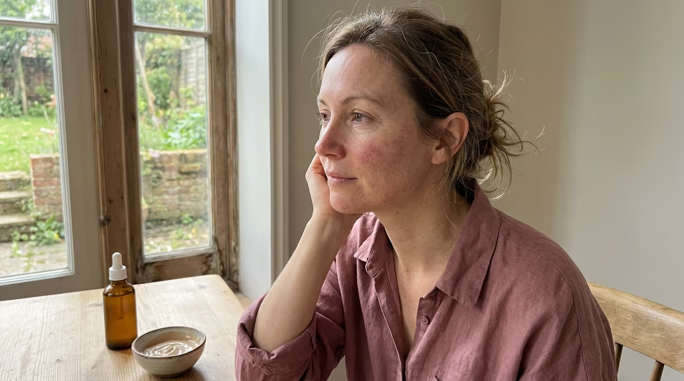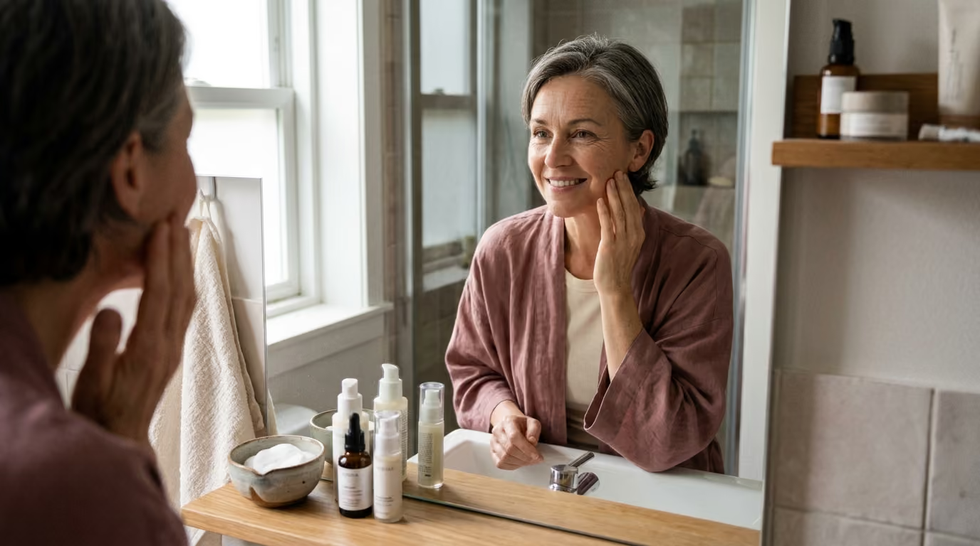

Chronic stress alters hormone signaling and weakens the skin barrier to accelerate visible aging across your face and body.





Chronic stress is not a personal failure or an imaginary state of mind. It is a measurable physiological state that influences hormones, immune signaling, nerve function, and cellular repair across the entire body. At the same time, chronic stress is not the sole cause of every sudden breakout, eczema patch, or fine line.
Skin changes occur through an intricate interplay of genetics, environment, immune health, sleep, and lifestyle. Viewing stress as one biological amplifier allows for practical solutions without placing blame on individuals dealing with complex skin concerns.
This guide examines the biological mechanisms connecting psychological strain to the cutaneous barrier, inflammatory cascades, wound repair, and visible skin aging. You will learn what clinical studies reveal, where current research reaches its limits, and how to create a calm, sustainable routine that protects your skin while supporting daily recovery.
To understand how prolonged pressure changes the skin, it helps to distinguish between acute and chronic strain. An acute stress response is a brief, adaptive reaction to an immediate demand. The body mobilizes energy, heightens awareness, and diverts resources toward immediate survival before returning to baseline balance.
Chronic stress occurs when psychological demands remain elevated over weeks, months, or years without adequate opportunity for recovery. The brain continuously perceives pressure, which drives persistent activation of two major neurological pathways: the sympathetic nervous system and the hypothalamic-pituitary-adrenal axis.
The sympathetic nervous system releases catecholamines, primarily epinephrine and norepinephrine. These chemical messengers travel through the bloodstream and bind directly to adrenergic receptors on blood vessels, immune cells, sebaceous glands, and epidermal keratinocytes. Constant sympathetic stimulation can constrict peripheral blood vessels, alter local nutrient delivery, and heighten sensory nerve sensitivity.
At the same time, the hypothalamic-pituitary-adrenal axis initiates a hormonal cascade. The hypothalamus releases corticotropin-releasing hormone, which prompts the pituitary gland to secrete adrenocorticotropic hormone. This signal travels to the adrenal glands, triggering the systemic release of cortisol.
Cortisol is essential for human life and helps regulate blood sugar, blood pressure, and inflammation. In short bursts, cortisol acts as an anti-inflammatory regulator. When elevation becomes prolonged or dysregulated, cortisol can alter immune responses, reduce the synthesis of key structural proteins, and interfere with normal tissue repair.
The connection between the mind and skin runs deeper than systemic blood flow. Research shows that human skin possesses its own fully functional, local neuroendocrine system. Keratinocytes, melanocytes, sebaceous cells, and dermal fibroblasts can synthesize corticotropin-releasing hormone, produce local glucocorticoids, and express receptors for these stress mediators.
Because the skin functions as a local stress organ, emotional strain can trigger cutaneous inflammation and barrier changes even when routine blood tests show normal systemic cortisol levels. Sensory nerves in the skin release neuropeptides such as substance P and calcitonin gene-related peptide when stimulated by mental distress. These neuropeptides interact with mast cells, prompting the release of histamine and inflammatory cytokines directly into surrounding tissue.
This local signaling network creates a continuous dialogue between the central nervous system and the cutaneous surface. Ongoing exposure to stress mediators alters the delicate balance between tissue destruction and renewal, laying the groundwork for heightened sensitivity, inflammatory flares, and compromised barrier integrity. For a broader look at cellular mechanisms, you can read our beauty science longevity guides on systemic cellular aging.
The stratum corneum is the outermost layer of the epidermis. It consists of protein-rich corneocytes surrounded by an organized lipid matrix made of ceramides, cholesterol, and free fatty acids. This structure keeps essential moisture inside while preventing environmental irritants, microbes, and allergens from penetrating deeper layers.
Scientific reviews show that sustained psychological stress impairs the production of both structural proteins and barrier lipids. When cortisol and catecholamines remain elevated, epidermal cells slow their production of filaggrin and loricrin. Filaggrin is a critical structural protein that aggregates keratin filaments and breaks down into natural moisturizing factors that maintain epidermal hydration.
At the same time, stress signaling reduces the activity of enzymes responsible for synthesizing ceramides and fatty acids in lamellar bodies. Without an adequate supply of intercellular lipids, the lipid bilayer becomes disorganized, creating microscopic gaps in the skin barrier.
This structural breakdown is measured clinically as transepidermal water loss. Transepidermal water loss measures the rate at which water vapor passes from inside the body through the epidermis into the surrounding atmosphere. Elevated water loss indicates that the physical barrier is compromised and struggling to retain moisture.
A weakened barrier produces noticeable daily symptoms:
When the barrier is thin and permeable, environmental irritants penetrate deeper into the viable epidermis. This irritation triggers the release of pro-inflammatory cytokines, creating mild, ongoing surface inflammation that further damages the barrier.
This dynamic explains why ordinary skincare can suddenly cause burning or redness during periods of high stress. The skin has not necessarily developed a permanent allergy to every ingredient. Instead, the physical defense system is temporarily compromised, allowing ingredients to reach sensitive nerve endings before the barrier can neutralize them.
Restoring barrier performance requires a gentle approach. When dealing with stress-induced barrier vulnerability, reducing active ingredients and focusing on basic moisture retention allows the stratum corneum time to rebuild its lipid bilayers. You can learn more about practical barrier support in our dedicated skin health and barrier care section.
Stress is widely recognized by dermatologists as a trigger that can aggravate pre-existing inflammatory skin conditions. The American Academy of Dermatology highlights that psychological strain frequently precedes or intensifies flares of acne, atopic dermatitis, psoriasis, rosacea, and chronic hives.
Stress does not create these conditions out of nowhere. Inflammatory skin diseases have complex roots involving genetics, immune tendencies, microbiome balance, and environmental triggers. Instead, stress functions as a biological amplifier that lowers the threshold for an inflammatory flare.
In acne-prone individuals, stress hormones stimulate sebaceous glands to produce more sebum. Corticotropin-releasing hormone can bind directly to sebocytes, increasing local oil production and promoting inflammatory signaling inside the hair follicle. Combined with altered follicular desquamation, this excess oil creates an ideal environment for Cutibacterium acnes proliferation, leading to painful inflammatory papules and pustules.
The relationship between stress and acne is also behavioral. During demanding periods, people often sleep fewer hours, change their nutritional habits, and forget their usual skincare routines. Many people also touch, pick, or squeeze blemishes unconsciously when feeling anxious. Repeated picking causes micro-trauma, introduces surface bacteria, and increases the risk of post-inflammatory hyperpigmentation and scarring.
For individuals with atopic dermatitis and eczema, stress triggers a debilitating itch-scratch cycle:
This cycle often peaks at night. As ambient distractions fade, the perception of itch increases, causing nighttime awakenings and restless sleep. The resulting sleep deprivation elevates systemic stress hormones the following day, making the skin even more reactive.
Psoriasis follows a similar neuro-immune pathway. Psychological stress stimulates the release of nerve growth factor, substance P, and inflammatory cytokines like interleukin-6 and tumor necrosis factor-alpha. These signaling molecules accelerate keratinocyte proliferation, leading to the rapid formation of thickened, scaly plaques.
Managing these flares requires addressing both the cutaneous inflammation and the underlying sensory signaling. Mind-body strategies, habit-reversal training, and consistent medical management work together to interrupt the cycle before permanent barrier damage occurs.
Wound healing is one of the most resource-intensive biological processes the body performs. Whether recovering from a minor scratch, an inflammatory breakout, or a cosmetic procedure, the skin relies on a coordinated sequence of cellular events.
Wound repair progresses through four distinct, overlapping phases:
Scientific reviews confirm that psychological stress can disrupt every phase of this repair process. Sustained elevations of cortisol and catecholamines impair the initial inflammatory response by altering cytokine recruitment. While excessive inflammation is harmful, a precise, controlled inflammatory burst is necessary to signal repair cells into the wounded area.
Prolonged stress signaling also impairs the proliferative phase. In laboratory studies on human skin cells, cortisol exposure significantly reduced the migratory capacity of keratinocytes. When keratinocytes cannot migrate efficiently, re-epithelialization slows, leaving the skin surface open to the environment for a longer period.
Dermal fibroblasts are equally vulnerable to stress hormones. Fibroblasts are responsible for manufacturing the structural proteins that support healing tissue. Under elevated cortisol conditions, fibroblast migration and proliferation decline, leading to delayed matrix deposition and weaker structural repair.
Stress also alters local tissue remodeling. Animal and human studies demonstrate that elevated glucocorticoid signaling suppresses the expression of growth factors, including fibroblast growth factor and vascular endothelial growth factor. Without adequate vascularization and matrix signaling, newly formed tissue lacks optimal resilience.
Behavioral factors further complicate tissue healing during stressful periods. Psychological strain frequently disrupts restorative sleep, during which human growth hormone and cellular repair pathways are most active. Poor nutritional intake, dehydration, smoking, and restless picking can all delay healing.
Understanding this biological reality helps set realistic expectations for skin recovery. If a blemish or minor procedure takes longer to resolve during an intense life phase, the delay reflects cellular resource allocation under strain rather than permanent damage.
The biological plausibility connecting chronic stress to visible skin changes has led researchers to investigate how emotional pressure affects the structural extracellular matrix over time. Chronic activation of stress pathways can increase oxidative stress, alter matrix metalloproteinase activity, and impair cellular repair mechanisms.
A landmark exploratory clinical study published in the Journal of Cosmetic Dermatology evaluated the physiological skin differences between women experiencing mild versus moderate chronic perceived stress. The study analyzed 36 healthy women aged 35 to 55 years, dividing them into two equal groups of 18 based on validated perceived-stress scales.
The researchers measured skin antioxidant status, barrier integrity, and high-resolution skin microtopography. The findings revealed statistically significant differences between the two groups:
To understand the biological mechanisms driving these structural differences, the investigators conducted laboratory experiments using cultured human skin cells and reconstructed human epidermis exposed to varying cortisol concentrations.
The cellular findings demonstrated direct effects on tissue building blocks:
These clinical and cellular measurements demonstrate that chronic psychological stress can influence parameters associated with skin barrier strength, antioxidant defense, and surface texture. However, these findings must be interpreted with nuance.
While the laboratory data explains how stress hormones interact with fibroblasts and keratinocytes, cellular experiments use isolated environments and fixed hormone concentrations. They confirm biological mechanisms, but they do not mean that an individual experiencing a difficult month will instantly develop deep structural wrinkles.
Visible skin changes develop over decades through cumulative factors including ultraviolet light exposure, intrinsic chronological aging, hormonal shifts, and lifestyle patterns. Chronic stress is best understood as one contributor to skin resilience rather than an unstoppable cause of visible aging. To read more about structural skin proteins, visit our guide on collagen and structural aging.
Scientific integrity requires understanding the boundaries of current research. While the connection between psychological stress and skin physiology is supported by numerous studies, the evidence base has specific limitations that prevent sweeping claims.
A major limitation in clinical psychodermatology is the distinction between correlation and causation. Observational studies can show that individuals with higher perceived stress have higher water loss or rougher skin texture. However, observation alone cannot prove that psychological stress directly caused those physical changes.
People under significant stress often experience lifestyle disruptions that independently influence skin health:
Disentangling the direct biological effects of cortisol from these secondary behavioral changes is exceptionally difficult in human clinical trials.
Sample sizes in dermatology stress research are often small. The previously cited clinical study comparing mild and moderate stress included 36 participants. While the methodology was rigorous, a cohort of 36 women aged 35 to 55 cannot represent the broader population. The study excluded men, smokers, individuals with poor sleep quality, and those with chronic medical conditions, limiting how broadly the findings apply.
Furthermore, translating laboratory cell-culture findings into real-world human outcomes presents major challenges. In-vitro experiments expose isolated fibroblasts or keratinocytes to constant, static concentrations of hydrocortisone or cortisol for 48 to 72 hours. In living humans, hormone secretion is pulsatile, follows a circadian rhythm, and interacts with dozens of buffering proteins and counter-regulatory hormones.
Measurement techniques for stress also vary across the scientific literature. Some researchers measure acute salivary cortisol, others test 24-hour urinary cortisol, and many rely exclusively on subjective perceived-stress questionnaires. Because systemic hormone levels do not always mirror local skin cortisol levels, comparing results across different trials can be inconsistent.
Finally, intervention studies evaluating mind-body therapies often face methodological hurdles. Clinical trials examining mindfulness, hypnotherapy, or relaxation techniques for eczema and psoriasis frequently suffer from small sample sizes, lack of blinded control groups, and short follow-up periods.
Mind-body interventions show promise as supportive tools, but the evidence does not support them as standalone replacements for established medical care. A balanced scientific perspective acknowledges stress as an important physiological factor without treating it as the sole determinant of skin health.
Navigating beauty advice requires separating evidence from common misconceptions. Marketing claims frequently oversimplify complex endocrinology, creating unnecessary worry for consumers.
Reality: Stress does not create acne on its own. Acne is a multifactorial inflammatory disease involving follicular hyperkeratinization, excess sebum, bacterial colonization, androgenic signaling, and genetic susceptibility. Stress hormones can aggravate existing pathways by increasing oil production and neurogenic inflammation, but emotional strain is not a personal failure that generates breakouts out of nothing.
Reality: There is no validated laboratory test that links a specific cortisol number to skin aging. Circulating cortisol fluctuates hourly based on circadian rhythms, meals, exercise, and immediate demands. Furthermore, because the skin produces its own local stress hormones, systemic lab tests cannot accurately reflect what is happening at the cellular level in your dermal fibroblasts.
Reality: Mind-body practices are valuable supportive tools, but they cannot replace evidence-based medical treatment. Conditions like severe eczema, plaque psoriasis, cystic acne, and rosacea involve complex immune pathways that usually require targeted topical therapies, systemic medications, barrier repair, and professional dermatologic care. Stress reduction supports the body while medical treatments address active disease.
Reality: While stress can heighten sensory nerve sensitivity and lower the itch threshold, persistent itching can signal underlying medical issues. Systemic conditions such as thyroid dysfunction, renal disease, hepatic changes, iron deficiency, or contact allergies can cause widespread pruritus. Unexplained, severe itching should always be evaluated by a healthcare professional rather than dismissed as stress.
Reality: When chronic stress compromises the epidermal barrier, the skin becomes more permeable and reactive. Introducing multiple new active ingredients like strong acids, high-strength retinoids, or complex botanical extracts during a high-stress period often causes contact irritation. A compromised barrier requires minimal, soothing ingredients to rebuild its lipid bilayers rather than intensive cosmetic intervention.
Supporting skin health during demanding periods does not require an elaborate routine. The goal is to reduce avoidable physiological demands on the skin barrier while creating realistic moments of nervous system recovery.
A practical approach uses the Protect, Treat, Recover model:
When life becomes demanding, simplify your topical routine to preserve the stratum corneum:
Do not abandon evidence-based dermatologic care in favor of stress-reduction techniques alone. Continue using prescribed topicals for acne, rosacea, or eczema as directed by your physician. Consistent medical management stabilizes inflammation, preventing the skin from sending distress signals back to the central nervous system.
Reducing physiological activation helps calm cutaneous sensory nerves and supports natural repair pathways. For broader recovery frameworks, explore our lifestyle recovery and environmental longevity resources.
Beneficial daily practices include:
If stress manifests as unconscious scratching or picking, use behavioral habit-reversal techniques:
Keeping a brief, objective log for two to three weeks can help you identify patterns without assigning blame. Track four simple metrics:
This objective data allows you to see how environmental shifts, sleep, and emotional demands interact, helping you adjust your care calmly and effectively.
To see how these concepts apply to everyday situations, review these common clinical patterns:
A 42-year-old professional experiences three weeks of intense project deadlines with reduced sleep. Her facial skin feels tight, dry, and stings when she applies her usual anti-aging serum.
Contributing factors:
Recommended response:
A 34-year-old teacher notices a cluster of deep, tender breakouts along her jawline during exam week. Anxious about the blemishes, she repeatedly picks at them in front of the mirror.
Contributing factors:
Recommended response:
A 48-year-old woman experiencing personal strain reports that her eczema has flared on her inner elbows and neck, with intense itching waking her at 3:00 AM.
Contributing factors:
Recommended response:
For a broader perspective on maintaining skin comfort and health over time, visit our comprehensive healthy aging longevity guide or review our evidence-based skin health resources.
No. An acute, short-term stressful event, such as giving a presentation or dealing with a brief scare, does not cause lasting skin damage or premature structural aging. The human body is well equipped to handle acute stress through adaptive neuroendocrine responses that quickly return to baseline.
Skin changes become noticeable only when psychological strain becomes chronic, persisting for months without adequate physical, emotional, or sleep recovery. Even under prolonged strain, the skin possesses significant regenerative capacity once supportive barrier care and restorative lifestyle habits are established.
Yes. The hair follicle is biologically sensitive to systemic and local neuroendocrine signaling. Severe or prolonged psychological stress can push a higher percentage of growing hair follicles prematurely into the resting phase, known as the telogen phase.
Approximately two to three months after the stressful period, individuals may notice increased daily shedding across the scalp, a condition called telogen effluvium. Like stress-related skin barrier disruption, telogen effluvium is usually temporary, and normal hair growth typically resumes once the underlying physiological demand resolves.
While mild dryness, temporary sensitivity, and minor stress breakouts can often be managed with gentle over-the-counter care, certain symptoms require professional medical evaluation.
You should consult a board-certified dermatologist or primary care physician if you experience:
Stay connected for research and practical guidance on skin, hair, collagen, nutrition and beauty longevity. Clear ideas for people who want to understand how appearance changes with age and make better-informed choices over time.


Understand your skin, hair and body better without chasing every new trend, treatment or promise.
explore the Blog