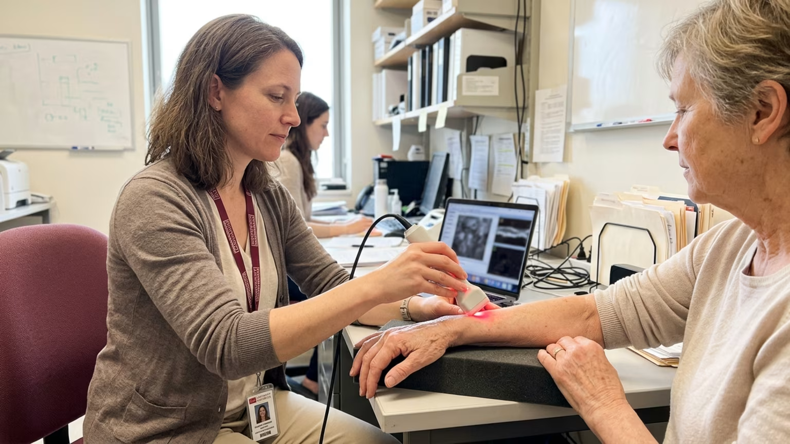

Boston University tested non-contact imaging for scleroderma skin changes. See the findings, study limits and why they do not establish a beauty test.





Boston University researchers tested a camera-based method for assessing skin changes in people with scleroderma, according to News-Medical’s October 14, 2026 report. The technique uses patterned light rather than physical contact to gather information from the skin.
Called spatial frequency domain imaging, or SFDI, the method produced readings associated with clinical skin scores, biopsy evidence of scarring and ultrasound-measured thickness. The study offers an early look at whether an optical measurement could help researchers and clinicians assess disease-related skin remodeling.
The researchers describe SFDI as projecting patterns of harmless light onto the skin and measuring the light that returns. A camera-based system records the signal without touching the skin. In this study, the team applied that approach to scleroderma, also known as systemic sclerosis.
The central research question was whether those light readings corresponded with other ways of assessing affected skin. The researchers compared SFDI with physician touch-based skin scores and ultrasound measurements of skin thickness. They also used a handheld measure of skin hardness and examined forearm biopsy findings.
These comparisons gave the team several reference points. The clinical assessment captured what a physician could feel during an examination. Ultrasound supplied a thickness measurement, while biopsies supplied tissue evidence of scarring and collagen buildup.
Corresponding author Andreea Bujor said the researchers examined these relationships alongside repeatability and changes over time. The study therefore considered both how the optical readings compared across participants and whether repeated readings might help follow skin changes.
According to News-Medical’s account, SFDI readings were lower in people with scleroderma than in healthy volunteers. Within the study, lower readings were associated with:
The finding is the alignment between the optical signal and several other assessments. The researchers were not simply showing that a camera could produce different readings from different people. They compared those readings with clinical findings and tissue-based evidence relevant to the disease.
The team also reported differences in SFDI readings in some areas that clinicians had scored as normal. That observation raises a research question about whether the optical signal captures changes that a standard examination does not register. It was an additional finding rather than the main comparison between affected participants and healthy volunteers.
A separate summary of the research described a study conducted at Boston Medical Center’s Scleroderma Clinic. It included 49 people with scleroderma and 25 healthy volunteers. Imaging covered six sites across the hands, fingers and forearms.
The researchers reported biopsy comparisons involving 34 patients and 16 healthy volunteers. Repeated measurements were conducted in 11 patients to examine whether SFDI readings tracked changes in clinical skin scores over time.
Those numbers describe different parts of the investigation. The full participant group supported the overall comparison. The biopsy group supported comparisons with tissue findings, while the repeat-measurement group addressed change over time.
These are participant counts, not diagnostic accuracy figures or treatment-success rates. As with research conducted under study conditions, performance in everyday clinical care may differ. The value of the counts is that they show the scale behind the reported findings rather than leaving “tested in patients” as an undefined claim.
The repeated-measurement evidence came from only 11 patients. That limits how broadly the findings about tracking change over time can be applied. Biopsy comparisons also involved fewer people than the full study cohort, so not every participant contributed tissue evidence.
The reported associations do not establish that SFDI directly measures collagen in routine use. They also do not establish that it diagnoses scleroderma, predicts patient outcomes or proves a response to treatment. A signal that corresponds with scarring is not automatically a validated test for every decision involving that scarring.
The available reporting does not provide sensitivity, specificity or accuracy figures. It also does not describe independent external validation. Without those results, the reports do not establish how reliably the method would distinguish relevant skin changes in a separate patient population.
The differences in clinically normal-scored skin need particular care in interpretation. They do not demonstrate that SFDI detects early scleroderma before a physician can. Establishing the clinical meaning of those differences would require further investigation.
Finally, the accounts provide summaries rather than the full original study results. They do not establish regulatory approval, commercial availability, reimbursement or readiness for routine practice. The supported interpretation is an assessment method under investigation, not a demonstrated replacement for examination, ultrasound or biopsy.
For readers interested in healthy aging, the practical lesson is to match a measurement claim to the condition actually studied. This research examined collagen-related fibrosis in scleroderma. It did not test cosmetic collagen supplements, topical products or interventions for ordinary skin aging.
Nothing in the reported results establishes a way to assess everyday age-related collagen loss or choose a personal skincare routine. The study therefore offers no basis for changing a supplement or topical product. Questions about monitoring scleroderma belong with a qualified clinician familiar with the person’s care.
Readers looking for broader background can turn to Younell’s guide to collagen within the skin’s supporting structure. Our resource on interpreting skin changes over time addresses the separate challenge of making sense of observations. Keeping disease research distinct from everyday beauty assessment helps prevent a promising measurement study from becoming an unsupported product claim.
Bujor said a fast, painless and non-contact measurement could help make skin monitoring more objective. She also described potential uses in clinical trials and in identifying subtle changes before they can be felt by hand.
The useful next question is where those optical readings add information that matters to patients and clinicians. Can changes in the signal consistently track meaningful changes in affected skin over time? Can the approach provide useful information alongside existing assessments?
For now, the study contributes a reported relationship between patterned-light readings and several signs of disease-related skin remodeling. Its significance lies in that measurement research. Further work will determine whether the relationship becomes a dependable tool for clinical monitoring.
Stay connected for research and practical guidance on skin, hair, collagen, nutrition and beauty longevity. Clear ideas for people who want to understand how appearance changes with age and make better-informed choices over time.





Understand your skin, hair and body better without chasing every new trend, treatment or promise.
explore the Blog