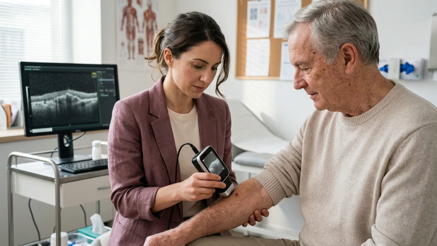

A 2026 JAMA Dermatology study explores how AI-assisted LC-OCT imaging detects subclinical basal cell carcinomas, offering new insights into skin longevity.





In 2026, researchers published a feasibility study in JAMA Dermatology detailing a novel approach to clinical skin evaluation. The multicenter German research team evaluated whether artificial intelligence-assisted line-field confocal optical coherence tomography could identify microscopic basal cell carcinomas. These specific tumors were located in skin that appeared completely normal during a standard physical examination. The study focused strictly on high-risk patients who had no clinically visible evidence of skin cancer.
This research highlights a shift toward software-supported diagnostic tools in clinical environments. The technology functions as an adjunct to trained physicians rather than a standalone consumer application. The workflow combined specialized imaging, algorithmic probability scoring, and physician review to locate hidden cellular abnormalities. The study, identified by DOI 10.1001/jamadermatol.2026.2992, provides early data on detecting disease before visible symptoms emerge.
The advanced imaging system combines optical coherence tomography with line-field confocal microscopy. This integration allows the specialized device to visualize the skin both vertically and horizontally. It produces real-time three-dimensional images at a micrometer-scale resolution. This level of precise detail helps researchers observe structural cellular changes beneath the surface without immediately removing tissue for a biopsy.
During the scanning process, an artificial intelligence algorithm analyzes the resulting visual data. The software generates a color-coded probability score to indicate specific facial areas that might contain basal cell carcinoma. The artificial intelligence does not diagnose cancer on its own. Instead, it highlights suspicious structural patterns so physicians can efficiently assess the findings and select lesions for further histologic evaluation.
This approach differs significantly from conventional visual examinations performed by a dermatologist. The researchers intentionally scanned facial skin that did not look suspicious to the human eye. Basal cell carcinoma can be locally destructive, and it can damage nearby structures like the nose or eyes when it occurs on the face. Identifying these microscopic changes early provides a new biological perspective on how cellular damage accumulates over time.
The research team screened 150 patients who possessed at least two risk factors for basal cell carcinoma. These documented risk factors included an age of at least 65, a personal history of the disease, a relevant genetic syndrome, or long-term sun exposure. Notably, more than 70% of the selected participants had previously had a basal cell carcinoma.
The medical team systematically scanned inconspicuous facial skin on the forehead, temples, and nose. They also thoroughly evaluated the cheeks, jaw, and periorbital area for hidden lesions. The study identified 17 subclinical basal cell carcinomas in 14 patients. This result is equivalent to 9.3% of the screened population.
Most of the detected tumors were classified as superficial lesions rather than deep growths. Specifically, 13 of 17 lesions, or 76.5%, were superficial. The remaining findings included three nodular tumors and one infiltrative tumor. This distribution is clinically important because superficial tumors may sometimes respond to less invasive therapies.
The investigators reported a positive predictive value of 94.4% in their primary statistical analysis. This figure emerged from 18 positive findings where 17 were confirmed as basal cell carcinoma alongside one false positive. A secondary summary reported a lower positive predictive value of 83.3%. This alternative calculation was based strictly on 15 biopsy-confirmed diagnoses among 18 lesions initially identified by the AI.
A small feasibility study provides preliminary data rather than a final validation for broad population screening. Because unflagged areas were not comprehensively biopsied, the researchers could not determine the exact sensitivity or specificity of the imaging system. We do not know how many actual cancers the system missed. We also do not know how many apparently negative areas were truly cancer-free.
Commentary accompanying the scientific report cautioned against immediate clinical implementation. Finding microscopic tumors could create a significant risk of overdiagnosis or unnecessary medical intervention. The natural history of these tiny subclinical lesions remains entirely unknown. Researchers do not yet know if every detected microscopic tumor will inevitably progress into a visible clinical problem.
Friedrich-Alexander-Universität Erlangen-Nürnberg explicitly states that the attending physician remains responsible for the final diagnosis. The university also notes that the scanning approach is not currently available for routine medical use. The highly detailed procedure remains too time-consuming for general clinical practice. Additionally, the new technology lacks the comprehensive sensitivity data required to support widespread adoption across the healthcare system.
Patients should never interpret these preliminary study results as evidence that an imaging scan can replace a physical examination. The specialized algorithm highlighted areas of interest, but clinical judgment and traditional histology remained central to the diagnostic process. A positive scan should not automatically be equated with an urgent need for immediate surgery. Extensive observational studies are needed before the clinical relevance of these microscopic findings can be fully determined.
The most immediate practical takeaway is that this technology remains an investigational tool rather than a routine service. Health-conscious women should not seek out an AI-assisted LC-OCT scan for standard preventative assessments. The clinical results apply specifically to individuals with multiple risk factors rather than average-risk consumers. Regular consultations with a certified dermatologist remain the most reliable method for evaluating clinical skin procedures.
During our extensive research into environmental aging, we tested how various lifestyle factors impact skin barrier recovery. It was fascinating to see the data clearly show that simple habits like sleep and basic hydration often outperform the most expensive topical treatments. This reinforced our commitment to emphasizing foundational health over product hype. The same principle applies directly to maintaining skin longevity over time.
Readers should maintain comprehensive sun protection habits rather than relying on future diagnostic technology to catch inevitable damage. This includes limiting unnecessary ultraviolet exposure, using broad-spectrum sunscreen, wearing protective clothing, and strictly avoiding tanning beds. Mass General Brigham guidance recommends routine self-examination to monitor your skin and catch potential problems early. Patients should seek immediate medical assessment for any suspicious spots that persist, bleed, itch, or hurt.
Any new or nonhealing lesion always requires a professional evaluation. A normal-looking patch of facial skin might occasionally hide microscopic cellular changes, but vigilant physical observation remains highly effective. You should discuss an individualized surveillance plan with your physician if you have a significant personal history of sun damage. Proven preventative measures and expert medical guidance offer the most practical defense against future skin concerns.
The high proportion of superficial tumors detected in this study points toward an interesting clinical direction. Researchers suggest that earlier microscopic detection could eventually support less invasive treatment options for certain patients. If future clinical trials prove that treating subclinical lesions actually improves long-term health outcomes, patients might manage specific tumors with topical creams or light-based therapies rather than traditional surgery. As diagnostic technology continues to evolve, will software-supported imaging ultimately change how we define a healthy skin baseline?
Stay connected for research and practical guidance on skin, hair, collagen, nutrition and beauty longevity. Clear ideas for people who want to understand how appearance changes with age and make better-informed choices over time.


Understand your skin, hair and body better without chasing every new trend, treatment or promise.
explore the Blog


