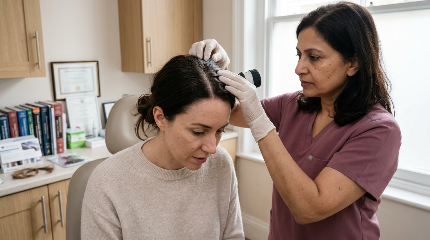

Three growth phases regulate follicular activity, guiding clinical intake, pull tests, trichoscopic imaging, and scalp evaluations to identify underlying.





When you first type "why is my hair falling out" into a search bar, the internet often presents an overwhelming list of catastrophic diagnoses, unproven home remedies, and aggressive marketing. Finding clumps of hair in the shower drain or noticing a widening part line can cause immediate distress. Many people spend months guessing at the cause, cycling through expensive shampoos and random dietary supplements with little success.
Diagnosing hair loss requires a methodical medical investigation rather than guesswork. A board-certified dermatologist or hair specialist approaches your scalp like a detective solving a biological puzzle. This comprehensive guide provides a definitive overview of how clinicians evaluate hair disorders, from the initial medical history to microscopic scalp imaging, targeted blood tests, and specialized biopsies.
Medical evaluation of the scalp follows an organized, evidence-based sequence. Rather than relying on a single test, clinicians combine non-invasive observations, bedside diagnostics, and laboratory tools to form an accurate diagnosis.
Hair loss is a symptom category rather than a single condition. A patient reporting that their ponytail feels thinner over several years has a distinct biological process from someone experiencing handfuls of shedding after a severe fever. Understanding the difference between shedding, thinning, and breakage helps guide the diagnostic journey.
Non-invasive methods form the backbone of routine clinical practice. Standardized global photography, systematic scalp inspection, hair-pull maneuvers, and trichoscopy allow clinicians to evaluate follicular health without causing tissue trauma. When these methods leave diagnostic ambiguity, semi-invasive and invasive tools offer deeper cellular clarity.
To understand diagnostic testing, one must understand the basic physiology of the hair growth cycle. Every hair follicle on the human scalp operates on an independent biological clock consisting of three primary phases. The anagen phase is the active growth period, lasting between two and seven years, during which matrix cells rapidly divide to construct the hair shaft.
The catagen phase represents a short transitional stage lasting approximately two weeks. During catagen, cell division ceases and the deeper portion of the follicle regresses toward the scalp surface. The telogen phase is the resting period, lasting roughly two to three months, where the club hair remains anchored in the follicle while cellular machinery resets.
At the conclusion of the telogen phase, the hair enters exogen, where the mature strand sheds from the scalp as a new anagen hair emerges beneath it. In a healthy scalp, approximately 85 to 90 percent of follicles remain in the anagen phase, while 10 to 15 percent rest in the telogen phase. Daily shedding of 50 to 100 telogen hairs reflects this standard cycle.
When physiological disruptions occur, this synchronized cycle shifts. In acute telogen effluvium, an abrupt physiological or metabolic shock triggers a large cohort of anagen hairs to exit active growth prematurely. Two to three months later, these hairs enter exogen simultaneously, producing alarming diffuse shedding. Because the follicular stem cell niche remains unharmed, this process does not involve miniaturization.
Progressive thinning involves an entirely different cellular mechanism known as follicular miniaturization. In androgenetic alopecia, genetically susceptible hair follicles contain higher levels of 5-alpha reductase and androgen receptors. When dihydrotestosterone binds to these receptors, it shortens the anagen phase and prolongs the telogen phase across successive cycles.
Over multiple cycles, the affected follicles produce progressively thinner, shorter, and less pigmented hair shafts. Eventually, terminal hairs transform into fine, vellus-like hairs that provide minimal scalp coverage. The total number of follicular openings may remain unchanged initially, but the reduction in average hair diameter creates visible thinning across the crown and frontal regions.
Scarring alopecias, or cicatricial alopecias, involve primary inflammatory destruction directed at the follicular stem cells located in the bulge region. Without viable stem cells, the follicle cannot generate a new anagen matrix. Fibrous scar tissue permanently replaces the follicular architecture, causing the scalp to lose its visible follicular openings.
Understanding these biological variations allows clinicians to look past superficial complaints. By examining whether a condition represents cycle synchronization, progressive miniaturization, or immune-mediated destruction, specialists can tailor their diagnostic protocols to the exact tissue layer involved. You can read more about foundational scalp science across our beauty science articles.
The medical history represents the most valuable diagnostic phase of a hair evaluation. A skilled practitioner spends substantial time analyzing the onset, tempo, and surrounding circumstances of your symptoms.
The clinician begins by establishing the exact timeline of your hair changes. They will ask whether the shedding began abruptly over a few days or evolved gradually over several years. They will also determine if the process is worsening, stabilizing, or fluctuating in cycles.
Sudden shedding that started three months after a severe illness points toward a reactive telogen shift. Conversely, a gradual reduction in density that has progressed quietly over five years suggests progressive pattern thinning. The clinician will ask where you notice shed hairs, whether in the shower, on your pillow, or across clothing.
They will also evaluate whether you notice localized bald patches, a widening midline part, or loss of body hair such as eyebrows and eyelashes. Scalp sensations provide critical diagnostic clues. Burning, itching, tenderness, or pain can signal active perifollicular inflammation, which warrants immediate investigation for scarring conditions.
Mapping where the hair loss occurs is essential. The clinician determines whether thinning involves the frontal hairline, the vertex crown, the temples, or the entire scalp uniformly. They will examine whether the occipital region at the lower back of the head remains dense and unaffected.
Family history must be thoroughly documented across both maternal and paternal lineages. A history of early pattern thinning among parents, grandparents, or siblings supports a diagnosis of androgenetic alopecia. A family history of autoimmune thyroid disease, vitiligo, or rheumatoid conditions raises the index of suspicion for alopecia areata or inflammatory scalp disorders.
Because hair follicles are among the most metabolically active tissues in the human body, they are sensitive to systemic shifts. The clinician will ask about events occurring two to six months prior to the onset of shedding. These triggers include high fevers, systemic infections, surgical procedures, general anesthesia, childbirth, and severe psychological stress.
Nutritional history receives close attention. Rapid weight loss, restrictive dieting, bariatric surgery, or inadequate dietary protein can disrupt normal follicular protein synthesis. Clinicians also review chronic medical symptoms such as persistent fatigue, cold sensitivity, unexplained weight fluctuations, joint stiffness, and menstrual irregularity.
Medication history is reviewed in detail. The clinician documents all prescription drugs, over-the-counter medications, herbal formulas, and high-dose dietary supplements. The start dates and dose adjustments of blood pressure drugs, antidepressants, hormonal therapies, oral retinoids, and cholesterol medications are compared against the hair loss timeline.
Mechanical tension and chemical processing can mimic or compound biological hair loss. Clinicians evaluate styling habits including tight ponytails, braids, weaves, extensions, and the use of chemical relaxers or permanent waves. Frequent thermal styling and aggressive brushing can cause extensive shaft fracture that patients mistake for primary shedding.
I remember speaking with a dermatologist who told me her patients were coming in with severe anxiety about normal skin aging. That anxiety was driven entirely by social media filters and aggressive marketing. That conversation became a cornerstone of our philosophy. We decided right then that our publication would never frame natural changes like wrinkles or thinning hair as personal failures.
Approaching your appointment with clarity and self-compassion helps you provide accurate clinical details without shame or panic. Learn more about our holistic, evidence-based approach on our about page.
Following the medical history, the specialist conducts a thorough physical inspection under focused, high-intensity lighting. The examination covers the entire scalp, hair shafts, nails, and other hair-bearing surfaces.
The practitioner methodically parts the hair in successive rows from the front of the scalp to the occiput. This systematic parting allows direct comparison between different anatomical zones. The clinician evaluates hair density, caliber uniformity, and the overall condition of the scalp skin.
During this visual inspection, the specialist checks whether follicular ostia, or openings, remain visible. The presence of normal follicular pores confirms that the underlying follicular structures are intact. In contrast, shiny, smooth, porcelain-like skin without follicular pores indicates that fibrous tissue has replaced the follicles.
The clinician also searches for signs of inflammation such as redness, scaling, pustules, crusting, or follicular plugging. Nail examination is routinely performed because systemic conditions and alopecia areata frequently produce nail abnormalities, such as pinpoint pitting, longitudinal ridging, or brittle splitting.
The hair-pull test is a standard bedside method used to estimate active follicular shedding. The clinician grasps a bundle of approximately 40 to 60 hair shafts between the thumb, index finger, and middle finger close to the scalp surface. They apply gentle, firm, slow traction along the entire shaft length away from the scalp.
This maneuver is repeated across multiple scalp regions, including the vertex, frontal zone, temporal areas, and occiput. In a normal scalp with stable cycling, no more than two to three hairs detach per pull site. Extracting more than 10 percent of the grasped bundle, roughly four to six hairs, indicates active shedding.
A positive hair-pull test confirms that an active shedding process is underway, but it does not diagnose the underlying cause. Brisk positive pulls can occur during acute telogen effluvium, active alopecia areata, or early inflammatory phases of scarring alopecia. Shed hairs can also be placed under a light microscope to evaluate the root bulb anatomy.
Microscopic evaluation distinguishes club-shaped telogen bulbs from tapered, dystrophic anagen roots. A negative pull test does not mean hair loss is absent. Patients with slow, chronic pattern thinning often exhibit negative pull tests because their follicles are miniaturizing rather than shedding rapidly.
Dermoscopy of the scalp, widely known as trichoscopy, has transformed hair diagnostics over the past two decades. Trichoscopy uses polarized and non-polarized magnification to visualize the epidermal surface, follicular ostia, perifollicular halos, and hair shaft architecture in real time.
Trichoscopy enables clinicians to identify microscopic signs that are invisible to the naked eye. In androgenetic alopecia, the hallmark feature is hair-diameter diversity exceeding 20 percent. While healthy scalps display uniform hair shaft thickness, miniaturizing scalps feature a mixture of thick terminal hairs, intermediate shafts, and fine vellus hairs emerging from the same region.
Another frequent sign in pattern loss is the peripilar sign, a subtle brownish or grayish halo around the follicular opening that reflects superficial perifollicular inflammation. The specialist may also observe empty follicular ostia, known as yellow dots, filled with sebum and keratin debris.
In alopecia areata, trichoscopy reveals distinct structural abnormalities caused by immune attacks on the hair matrix. Black dots represent shafts that have fractured at the level of the scalp surface. Exclamation-mark hairs are short, broken hairs whose proximal ends near the scalp are distinctly thinner than their distal tips.
In telogen effluvium, trichoscopy often reveals normal hair-shaft diameters without significant caliber diversity. Clinicians typically observe short, upright regrowing hairs emerging from normal follicular openings several weeks after the primary trigger resolves. Because specific positive markers are scarce, telogen effluvium often serves as a trichoscopic diagnosis of exclusion.
In scarring alopecias such as lichen planopilaris and frontal fibrosing alopecia, trichoscopy detects the complete absence of follicular openings in affected zones. Clinicians look for tubular perifollicular scaling, bright erythema, and tufted hairs where multiple shafts emerge from a single fibrotic opening. You can find more practical frameworks in our hair health resources.
Medical research supports the diagnostic accuracy of trichoscopy in distinguishing between complex hair disorders. In a clinical study published in the International Journal of Trichology, researchers evaluated 21 complex alopecia cases and found that trichoscopy established a definitive diagnosis in 19 cases, representing a 90.5 percent diagnostic success rate without requiring initial invasive procedures.
A systematic review published in the Journal of the European Academy of Dermatology and Venereology examined the diagnostic metrics of trichoscopic features in pattern hair loss. The meta-analysis established that hair-diameter diversity greater than 20 percent exhibited a sensitivity of 94.07 percent and a positive predictive value of 95.99 percent for diagnosing androgenetic alopecia.
Research evaluating female pattern hair loss established that combining specific major criteria, such as frontal yellow dots and decreased average thickness, with minor criteria yielded a diagnostic specificity of 92 percent. These quantitative metrics demonstrate that magnified scalp examination provides objective, reproducible data that substantially reduces diagnostic uncertainty.
While modern diagnostic tools are powerful, they have inherent limitations that clinicians and patients must recognize. No single bedside test or trichoscopic sign is completely infallible on its own.
A prime example is the negative predictive value of hair-diameter diversity. While diameter variation carries a positive predictive value of nearly 96 percent in pattern loss, its negative predictive value was documented at only 30.76 percent in systematic reviews. This low negative predictive value means that the absence of visible diameter diversity on a single examination cannot completely rule out early androgenetic thinning.
The hair-pull test is also subject to substantial operational variability. If a patient shampoos vigorously immediately prior to their appointment, they may wash away loose telogen hairs, producing a false-negative test result. Conversely, if a patient avoids washing their hair for a week due to shedding anxiety, the accumulation of normal shed hairs can cause an exaggerated false-positive pull test.
Trichoscopy also has diagnostic boundaries. While it excels at identifying active inflammation and shaft variations, it cannot evaluate cellular changes deep within the reticular dermis or subcutaneous fat. In early scarring alopecia, surface signs can be subtle and easily confused with severe seborrheic dermatitis or chronic pattern loss.
Relying entirely on consumer scalp cameras or automated smartphone applications presents significant risks. Without formal dermatologic training, patients often misinterpret normal anatomical landmarks as pathological signs, generating unnecessary stress. Trichoscopic findings must always be interpreted alongside the patient's complete history and physical exam.
When history and physical examination leave diagnostic questions or point toward systemic factors, clinicians turn to targeted laboratory panels and tissue biopsies.
Blood tests are not ordered indiscriminately for every patient. Instead, testing is tailored to the individual clinical presentation, demographic profile, and suspected pathology.
Ferritin is particularly important because it measures intracellular iron storage. While a ferritin level of 15 nanograms per milliliter may be considered clinically normal for general health, hair follicles require adequate iron stores to support rapid matrix cell division. Thyroid-stimulating hormone testing identifies subtle hypothyroidism or hyperthyroidism, both of which can disrupt the follicular cycle.
When female patients exhibit signs of hyperandrogenism, such as irregular menses, severe adult acne, or hirsutism, clinicians may order free and total testosterone, androstenedione, and DHEA-sulfate. However, most women with female pattern hair loss exhibit normal circulating androgen levels, as their condition stems from heightened follicular sensitivity rather than systemic hormonal excess.
Normal blood tests do not mean your hair loss is imaginary. Common conditions such as androgenetic alopecia, alopecia areata, traction alopecia, and early scarring disorders routinely present with completely normal laboratory values. Blood testing is designed to identify systemic contributors, not to serve as a standalone test for primary hair loss.
A scalp biopsy is the definitive diagnostic procedure when scarring alopecia is suspected or when non-invasive tools fail to establish a clear diagnosis. It is a minor in-office surgical procedure performed under local anesthesia.
The practitioner selects an active, hair-bearing margin of the affected scalp. Choosing a completely bald, smooth area is a common diagnostic mistake. In end-stage scar tissue, the follicular structures have already vanished, leaving the pathologist with little active tissue to evaluate.
The clinician typically obtains two 4-millimeter punch biopsies taken parallel to the natural angle of the hair shafts. The punch must penetrate through the dermis and reach the subcutaneous fat layer, where anagen hair bulbs reside. The biopsy sites are closed with small sutures that are removed 7 to 10 days later.
Standard pathology protocol processes one specimen using horizontal cross-sections and the other using vertical longitudinal sections. Horizontal sectioning allows the dermatopathologist to count the total number of follicles, determine the ratio of terminal to vellus hairs, and calculate the exact percentage of follicles in anagen versus telogen.
Vertical sectioning allows detailed visualization of the dermal-epidermal junction, the follicular infundibulum, and inflammatory cell patterns around the upper bulge area. This dual-specimen approach provides the structural and cellular clarity required to distinguish between complex conditions such as lichen planopilaris, central centrifugal cicatricial alopecia, and lupus erythematosus. To browse related long-form guides, visit our complete research library.
Misunderstandings surrounding hair loss testing can lead to misdirected treatments and unwarranted anxiety. Clarifying these issues helps establish realistic expectations for clinical appointments.
Many patients believe that if hairs come out easily during a pull test, those follicles are permanently dead. In reality, a positive pull test simply reflects hairs transitioning through the telogen phase. Conditions like acute telogen effluvium can produce alarming pull test results while preserving the underlying follicular machinery for complete recovery.
Patients are often told by non-specialists that because their blood count and thyroid levels are normal, their hair thinning is purely cosmetic or imaginary. As clinical data demonstrates, androgenetic alopecia and early cicatricial conditions develop locally at the scalp tissue level without altering circulating blood markers. A normal lab panel is reassurance that systemic illness is absent, not proof of healthy follicles.
Digital USB microscopes marketed directly to consumers are widely available online. While these devices can show magnified views of the scalp surface, capturing an image is very different from interpreting complex biological architecture. Without clinical context, patients frequently mistake benign sebum plugs for disease or overlook subtle perifollicular erythema that signals early scarring.
Shedding and thinning are distinct biological processes that require different diagnostic approaches. Shedding refers to an increased daily loss of hair strands with club roots, reflecting altered cycle dynamics. Thinning refers to the progressive reduction in hair shaft diameter due to follicular miniaturization. A patient can experience progressive pattern thinning with minimal daily shedding, or heavy daily shedding without long-term density loss.
Washing your hair immediately before an appointment can wash away loose telogen hairs, which temporarily masks active shedding during a physical pull test. Most dermatologists recommend washing your scalp 24 to 48 hours before your visit. This interval allows natural shedding patterns to remain observable during the bedside exam without causing excessive sebum buildup that could obscure trichoscopy.
A standard 4-millimeter scalp biopsy creates a small circular incision that is closed with one or two sutures. The superficial skin layer typically closes within 7 to 10 days, at which point the sutures are removed. Because the biopsy removes hair follicles at that specific site, it leaves a tiny, pencil-tip-sized scar that is easily hidden by surrounding hair.
Yes, overlapping hair disorders are common in clinical practice. A patient can have underlying, slowly progressive androgenetic alopecia and subsequently develop acute telogen effluvium following a major surgery or illness. Similarly, it is possible for a patient to have both traction alopecia along the frontal hairline and an inflammatory condition on the crown. A thorough diagnostic evaluation identifies each contributing component.
While hair density naturally decreases slightly over decades, noticeable or rapid thinning is not something you must dismiss without evaluation. If a general practitioner cannot provide a clear structural explanation for your symptoms, request a referral to a board-certified dermatologist who specializes in hair and scalp disorders. Specialized tools like trichoscopy can distinguish normal age-related changes from treatable medical conditions.
There is no routine blood test that diagnoses genetic pattern hair loss. Genetic hair loss is diagnosed through clinical examination, family history, and trichoscopic evidence of hair-diameter diversity. While commercial genetic swab tests exist, they offer limited clinical utility because genetic susceptibility involves multiple interactive genes rather than a single measurable marker.
A productive diagnostic appointment requires thoughtful preparation. Going into your appointment organized helps your specialist gather accurate clinical data and form an effective management plan.
Reviewing your daily routine before your visit helps you provide clear answers during your consultation. You can learn more about how nutrition, lifestyle, and hair health intersect in our hair health section.
Stay connected for research and practical guidance on skin, hair, collagen, nutrition and beauty longevity. Clear ideas for people who want to understand how appearance changes with age and make better-informed choices over time.


Understand your skin, hair and body better without chasing every new trend, treatment or promise.
explore the Blog